Ito M, Oishi R, Fukunaga M, Sone T, Sugimoto T, Shiraki M, Nishizawa Y, Nakamura T
Medical Work-Life-Balance Center, Nagasaki University Hospital, 1-7-1 Sakamoto, Nagasaki, 852-8501, Japan,
Osteoporos Int. 2014 Mar;25(3):1163-72. doi: 10.1007/s00198-013-2596-y. Epub 2013 Dec 18.
Once-weekly administration of 56.5 μg teriparatide improved cortical bone parameters and biomechanical parameters at the proximal femur by CT geometry analysis.
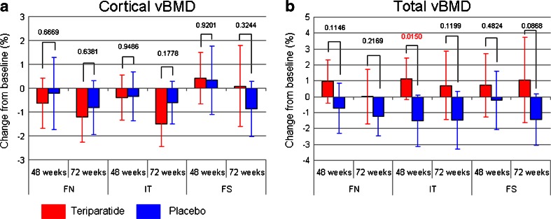
The aim of this study was to evaluate the effects of weekly administration of teriparatide [human PTH (1-34)] on bone geometry, volumetric bone mineral density (vBMD), and parameters of bone strength at the proximal femur which were longitudinally investigated using computed tomography (CT).
The subjects were a subgroup of a recent, randomly assigned, double-blind study (578 subjects) comparing the anti-fracture efficacy of a once-weekly subcutaneous injection of 56.5 μg teriparatide with placebo (TOWER trial).
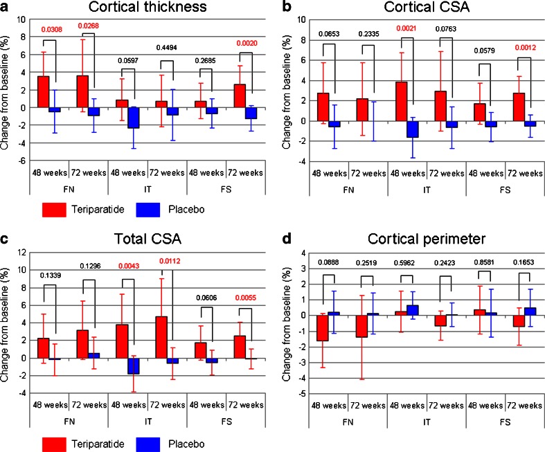
Sixty-six ambulatory postmenopausal women with osteoporosis were enrolled at 15 study sites having multi-detector row CT, and included women injected with teriparatide (n = 29, 74.2 ± 5.1 years) or with placebo (n = 37, 74.8 ± 5.3 years). CT data were obtained at baseline and follow-up scans were performed at 48 and 72 weeks. The data were analyzed to obtain cross-sectional densitometric, geometric, and biomechanical parameters including the section modulus (SM) and buckling ratio (BR) of the femoral neck, inter-trochanter, and femoral shaft. We found that once-weekly teriparatide increased cortical thickness/cross-sectional area (CSA) and total area, and improved biomechanical properties (i.e., decreasing BR) at the femoral neck and shaft. Teriparatide did not change the cortical perimeter.
Our longitudinal analysis of proximal femur geometry by CT revealed that once-weekly administration of 56.5 μg teriparatide improved cortical bone parameters at the femoral neck and shaft and also improved biomechanical parameters.
通过CT几何分析,每周一次给予56.5μg特立帕肽可改善股骨近端的皮质骨参数和生物力学参数。
本研究的目的是评估每周给予特立帕肽[人甲状旁腺激素(1-34)]对骨几何形态、骨体积密度(vBMD)以及使用计算机断层扫描(CT)纵向研究的股骨近端骨强度参数的影响。
受试者是最近一项随机分配的双盲研究(578名受试者)的一个亚组,该研究比较了每周一次皮下注射56.5μg特立帕肽与安慰剂的抗骨折疗效(TOWER试验)。
66名患有骨质疏松症的绝经后门诊妇女在15个拥有多排探测器CT的研究地点入组,包括注射特立帕肽的妇女(n = 29,74.2±5.1岁)或注射安慰剂的妇女(n = 37,74.8±5.3岁)。在基线时获取CT数据,并在48周和72周进行随访扫描。对数据进行分析以获得横断面密度测量、几何和生物力学参数,包括股骨颈、转子间和股骨干的截面模量(SM)和屈曲比(BR)。我们发现每周一次的特立帕肽增加了皮质厚度/横截面积(CSA)和总面积,并改善了股骨颈和骨干的生物力学性能(即降低BR)。特立帕肽未改变皮质周长。
我们通过CT对股骨近端几何形态的纵向分析表明,每周一次给予56.5μg特立帕肽可改善股骨颈和骨干的皮质骨参数,并且还改善了生物力学参数。