Kyadari Mahender, Fatma Tasneem, Azad Rajvardhan, Velpandian Thirumurthy
Department of Pharmacy, Integrated Institute of Technology, Dwarka, New Delhi, India.
Department of Bio-Sciences, Jamia Millia Islamia, New Delhi, India.
Indian J Pharmacol. 2013 Nov-Dec;45(6):569-74. doi: 10.4103/0253-7613.121366.
algae isolates obtained from fresh and marine resources could be one of the richest sources of novel bioactive secondary metabolites expected to have pharmaceutical significance for new drug development. This study was conducted to evaluate the antiangiogenic and antiproliferative activity of Chlorella pyrenoidosa in experimental models of angiogenesis and by MTT assay.
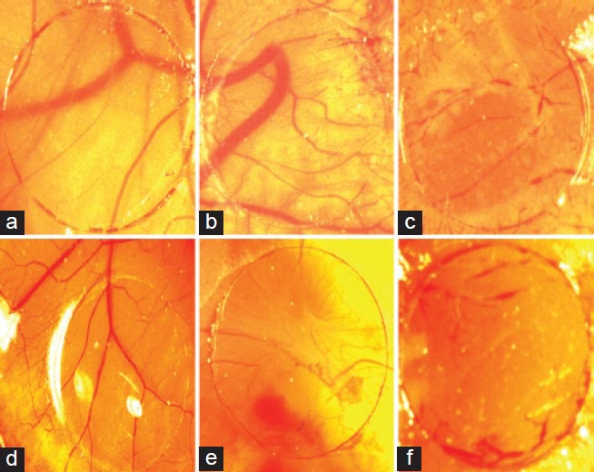
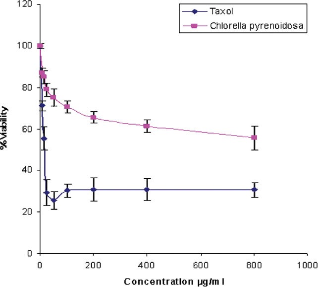
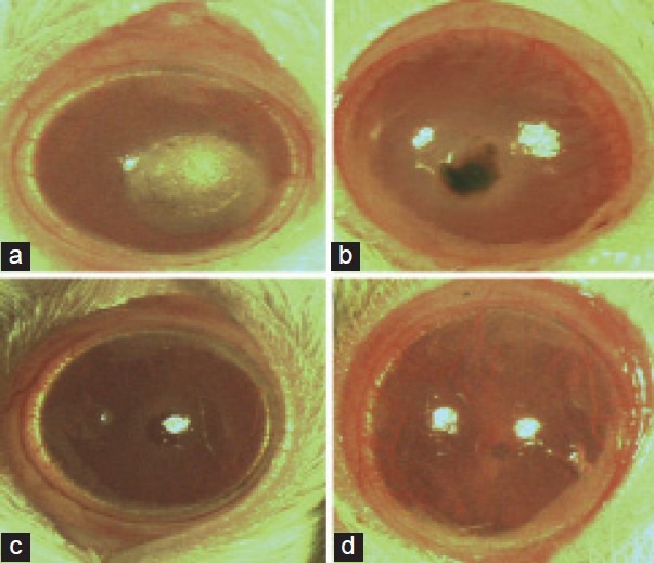
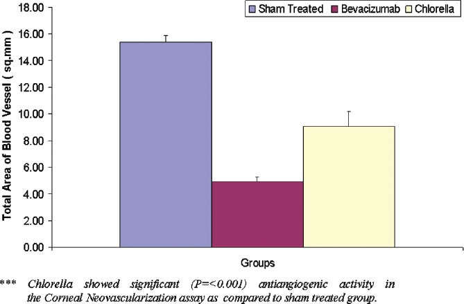
lyophilized extract of C. pyrenoidosa was extracted using dichloromethane/methanol (2:1), concentrated and vacuum evaporated to obtain the dried extract. The crude extract was evaluated in the vascular endothelial growth factor (VEGF)-induced angiogenesis in in ovo chick chorioallantoic membrane assay (CAM) at various concentrations (n = 8) using thalidomide and normal saline as positive and untreated control groups, respectively. The crude extract was also subjected to the antiangiogenic activity in the silver nitrate/potassium nitrate cautery model of corneal neovascularization (CN) in rats where topical bevacizumab was used as a positive control. The vasculature was photographed and blood vessel density was quantified using Aphelion imaging software. The extract was also evaluated for its anti proliferative activity by microculture tetrazolium test (MTT) assay using HeLa cancer cell line (ATCC).
VEGF increased the blood vessel density by 220% as compared to normal and thalidomide treatment decreased it to 67.2% in in ovo assay. In the in-vivo CN model, the mean neovascular density in the control group, the C. pyrenoidosa extract and bevacizumab group were found to be 100%, 59.02%, and 32.20%, respectively. The Chlorella pyrenoidosa extract negatively affected the viability of HeLa cells. An IC50 value of the extract was 570 μg/ml, respectively.
a significant antiangiogenic activity was observed against VEGF-induced neovascularization and antiproliferative activity by MTT assay. In this study, it could be attributed that the activity may be due to the presence of secondary metabolites in the C. pyrenoidosa extract.
从淡水和海洋资源中分离得到的藻类可能是新型生物活性次生代谢产物最丰富的来源之一,有望对新药开发具有药学意义。本研究旨在通过血管生成实验模型和MTT法评估小球藻的抗血管生成和抗增殖活性。
采用二氯甲烷/甲醇(2:1)提取小球藻冻干提取物,浓缩并真空蒸发得到干燥提取物。在不同浓度(n = 8)下,分别以沙利度胺和生理盐水作为阳性对照组和未处理对照组,通过鸡胚绒毛尿囊膜试验(CAM)评估粗提物对血管内皮生长因子(VEGF)诱导的血管生成的影响。粗提物还在大鼠角膜新生血管(CN)的硝酸银/硝酸钾烧灼模型中进行抗血管生成活性研究,局部使用贝伐单抗作为阳性对照。对血管系统进行拍照,并使用Aphelion成像软件对血管密度进行定量分析。还采用HeLa癌细胞系(ATCC)通过微量培养四氮唑蓝试验(MTT)评估提取物的抗增殖活性。
在鸡胚试验中,与正常情况相比,VEGF使血管密度增加了220%,而沙利度胺处理使其降至67.2%。在体内CN模型中,对照组、小球藻提取物组和贝伐单抗组的平均新生血管密度分别为100%、59.02%和32.20%。小球藻提取物对HeLa细胞的活力有负面影响。提取物的IC50值分别为570μg/ml。
通过MTT法观察到小球藻提取物对VEGF诱导的新生血管生成具有显著的抗血管生成活性和抗增殖活性。在本研究中,该活性可能归因于小球藻提取物中存在的次生代谢产物。