Barchetti F, De Marco V, Barchetti G, Pasqualitto E, Sartori A, Glorioso M, Gigli S, Megna V, Montechiarello S, Boncore V, Stagnitti A
Department of Radiological Sciences, Oncology and Pathology, Sapienza University of Rome, Rome, Italy.
Case Rep Oncol. 2013 Oct 24;6(3):520-5. doi: 10.1159/000356097. eCollection 2013.
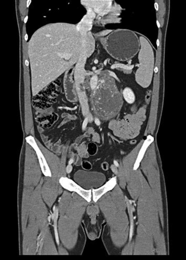
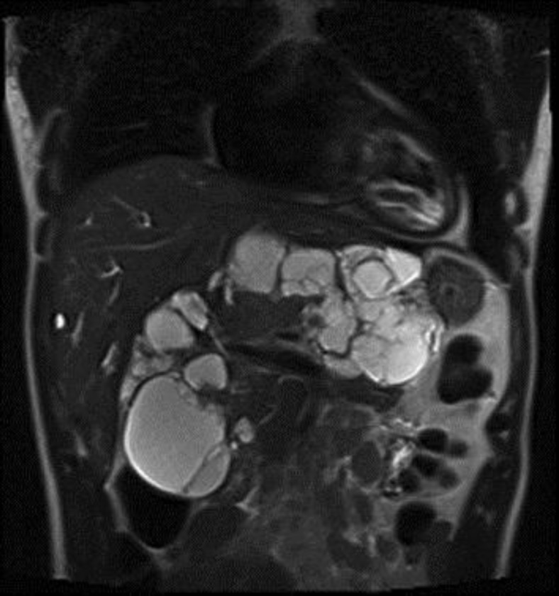
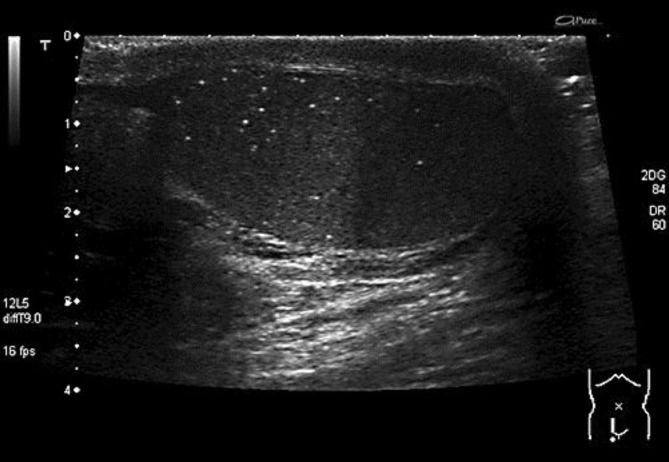
Many studies have demonstrated an association between diffuse bilateral testicular microlithiasis (TM) and gonadal and extragonadal germ cell tumors. Nevertheless, it is still uncertain whether ultrasound surveillance is really necessary in patients with TM in the absence of other risk factors such as previous testicular cancer, a history of cryptorchidism or testicular atrophy. We report the cases of a 33- and a 39-year-old man presenting with a retroperitoneal extragonadal tumor. The first patient underwent an MRI examination in order to rule out a lumbosacral hernia: MRI images showed no slipped disks but a voluminous retroperitoneal solid mass. The histological analysis revealed an immature teratoma. The second patient came to the emergency department complaining of abdominal pain, vomiting, weight loss and mild jaundice: ultrasound examination showed a large, ill-defined heterogeneous abdominal mass, confirmed by CT and MRI examination. The histology diagnosed a yolk sac tumor. In both patients, the testicular sonography was performed to rule out a focal lesion, but it displayed bilateral TM without a focal testicular mass. Based on our direct experience, we highlight the importance of annual ultrasonographic surveillance of the testis and the retroperitoneal space in patients with occasionally detected TM.
许多研究已证实弥漫性双侧睾丸微结石症(TM)与性腺及性腺外生殖细胞肿瘤之间存在关联。然而,在没有其他危险因素(如既往睾丸癌、隐睾病史或睾丸萎缩)的TM患者中,超声监测是否真的必要仍不确定。我们报告了一名33岁和一名39岁男性出现腹膜后性腺外肿瘤的病例。第一名患者接受了MRI检查以排除腰骶部疝:MRI图像显示无椎间盘突出,但有一个巨大的腹膜后实性肿块。组织学分析显示为未成熟畸胎瘤。第二名患者因腹痛、呕吐、体重减轻和轻度黄疸到急诊科就诊:超声检查显示一个大的、边界不清的异质性腹部肿块,CT和MRI检查证实。组织学诊断为卵黄囊瘤。在这两名患者中,均进行了睾丸超声检查以排除局灶性病变,但显示双侧TM且无睾丸局灶性肿块。基于我们的直接经验,我们强调对于偶尔发现TM的患者,每年对睾丸和腹膜后间隙进行超声监测的重要性。