Iurilli Giuliano, Olcese Umberto, Medini Paolo
Department of Neuroscience and Brain Technologies, Istituto Italiano di Tecnologia, Genova, Italy.
PLoS One. 2013 Dec 12;8(12):e82044. doi: 10.1371/journal.pone.0082044. eCollection 2013.
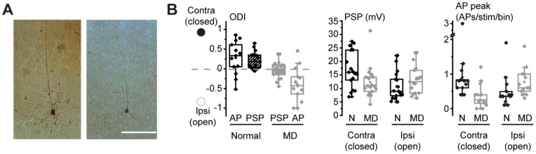
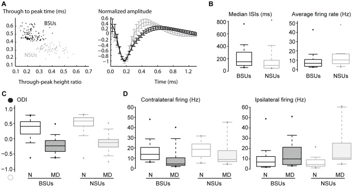
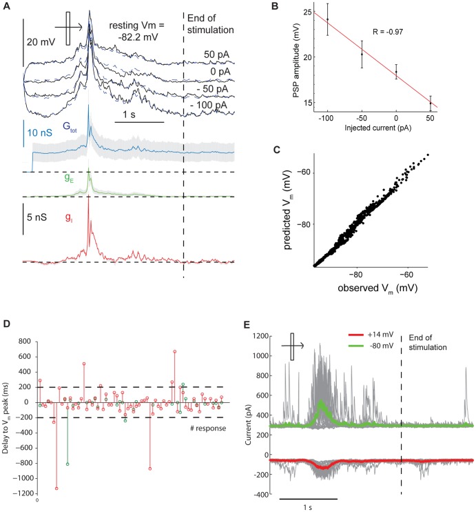
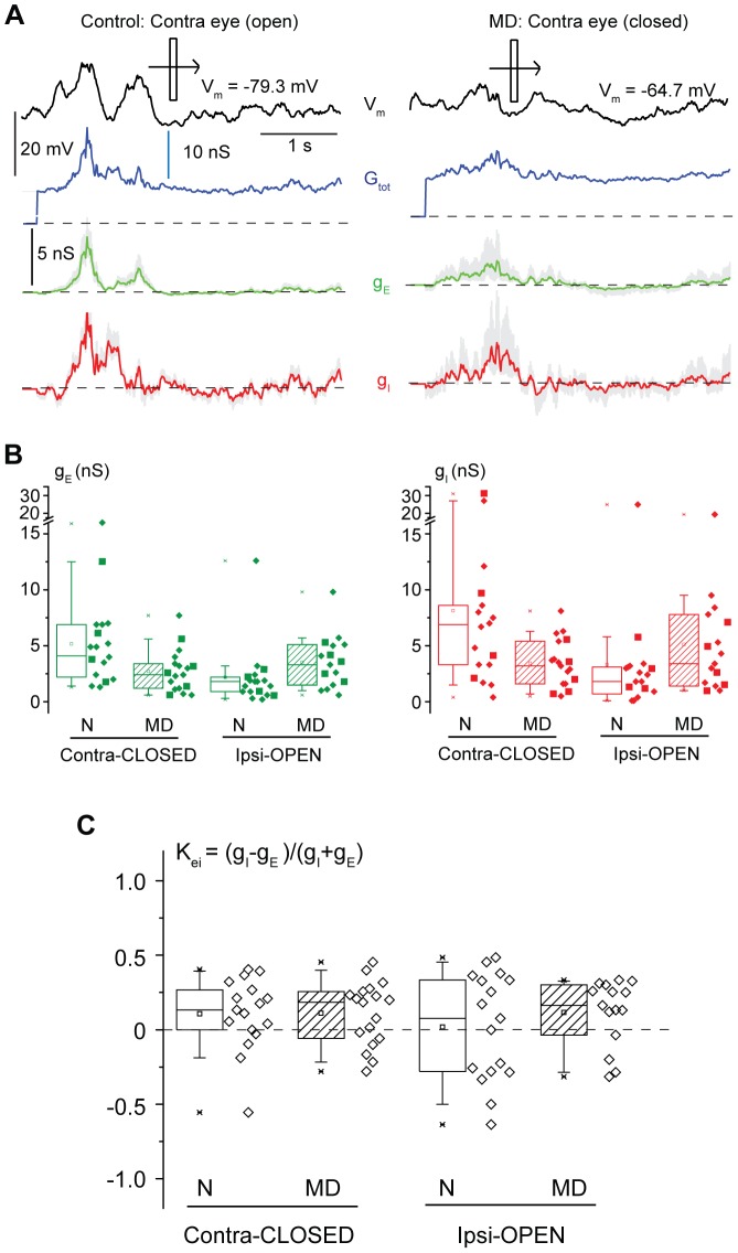
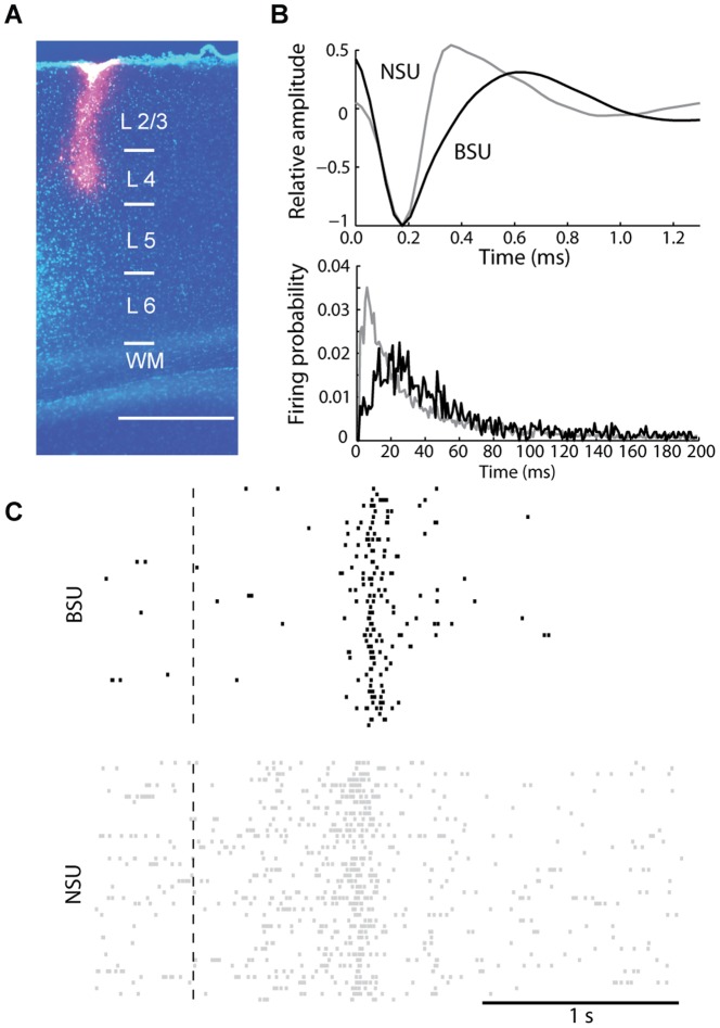
Monocular deprivation (MD) during development leads to a dramatic loss of responsiveness through the deprived eye in primary visual cortical neurons, and to degraded spatial vision (amblyopia) in all species tested so far, including rodents. Such loss of responsiveness is accompanied since the beginning by a decreased excitatory drive from the thalamo-cortical inputs. However, in the thalamorecipient layer 4, inhibitory interneurons are initially unaffected by MD and their synapses onto pyramidal cells potentiate. It remains controversial whether ocular dominance plasticity similarly or differentially affects the excitatory and inhibitory synaptic conductances driven by visual stimulation of the deprived eye and impinging onto visual cortical pyramids, after a saturating period of MD. To address this issue, we isolated visually-driven excitatory and inhibitory conductances by in vivo whole-cell recordings from layer 4 regular-spiking neurons in the primary visual cortex (V1) of juvenile rats. We found that a saturating period of MD comparably reduced visually-driven excitatory and inhibitory conductances driven by visual stimulation of the deprived eye. Also, the excitatory and inhibitory conductances underlying the synaptic responses driven by the ipsilateral, left open eye were similarly potentiated compared to controls. Multiunit recordings in layer 4 followed by spike sorting indicated that the suprathreshold loss of responsiveness and the MD-driven ocular preference shifts were similar for narrow spiking, putative inhibitory neurons and broad spiking, putative excitatory neurons. Thus, by the time the plastic response has reached a plateau, inhibitory circuits adjust to preserve the normal balance between excitation and inhibition in the cortical network of the main thalamorecipient layer.
发育过程中的单眼剥夺(MD)会导致初级视皮层神经元中被剥夺眼的反应性急剧丧失,并且在包括啮齿动物在内的迄今所有测试物种中都会导致空间视觉退化(弱视)。这种反应性丧失从一开始就伴随着丘脑 - 皮质输入的兴奋性驱动降低。然而,在丘脑接受层4中,抑制性中间神经元最初不受MD影响,并且它们与锥体细胞的突触增强。在MD达到饱和期后,眼优势可塑性是否同样或不同地影响由被剥夺眼的视觉刺激驱动并作用于视觉皮质锥体的兴奋性和抑制性突触电导,这仍然存在争议。为了解决这个问题,我们通过在幼年大鼠初级视皮层(V1)的第4层规则发放神经元进行体内全细胞记录,分离出视觉驱动的兴奋性和抑制性电导。我们发现,MD的饱和期同样降低了由被剥夺眼的视觉刺激驱动的视觉驱动兴奋性和抑制性电导。此外,与对照组相比,由同侧未被剥夺的左眼驱动的突触反应所基于的兴奋性和抑制性电导同样增强。在第4层进行多单元记录并随后进行尖峰分类表明,对于窄峰发放的假定抑制性神经元和宽峰发放的假定兴奋性神经元,反应性的阈上丧失和MD驱动的眼偏好转移是相似的。因此,当可塑性反应达到平稳期时,抑制性回路会进行调整,以维持主要丘脑接受层皮质网络中兴奋和抑制之间的正常平衡。