Maternal and Fetal Health Research Centre, Institute of Human Development, University of Manchester, Manchester Academic Health Sciences Centre, St Marys Hospital, Manchester M13 9WL, UK.
Mol Hum Reprod. 2014 May;20(5):433-41. doi: 10.1093/molehr/gat095. Epub 2013 Dec 19.
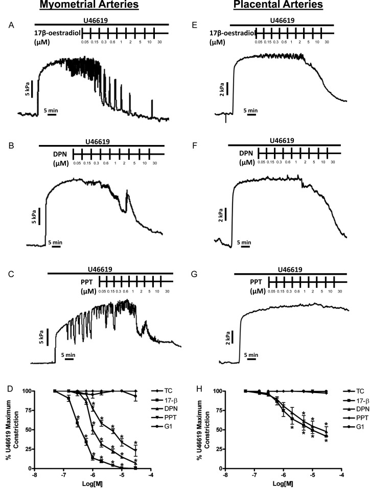
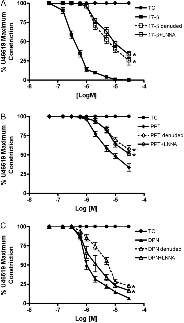
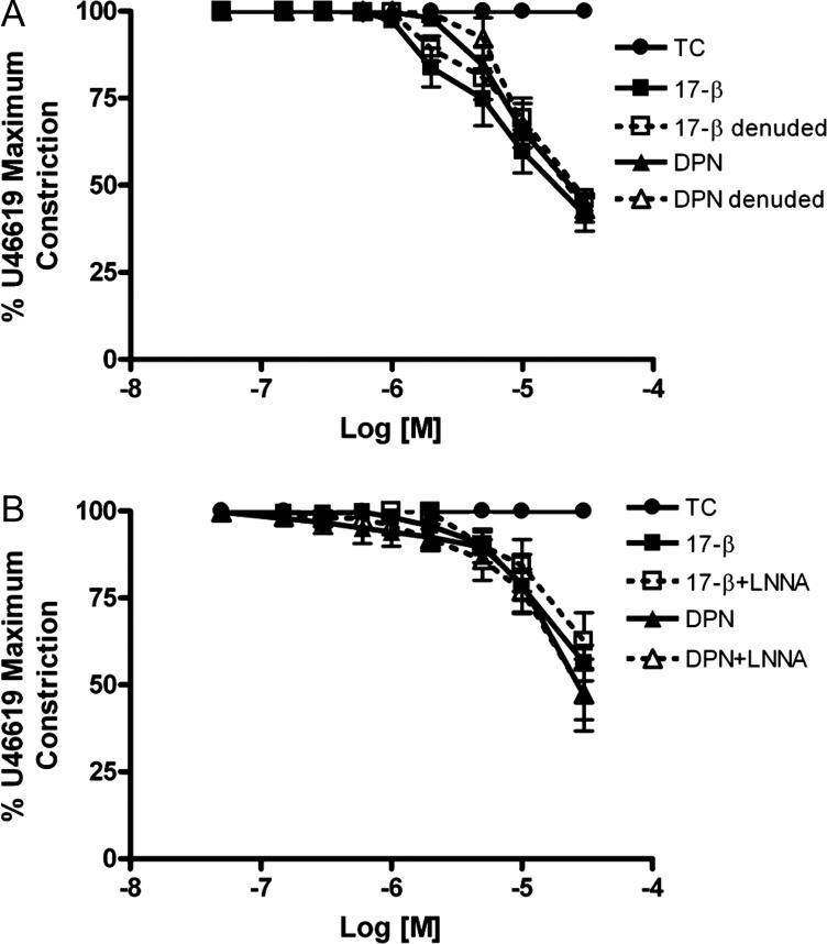
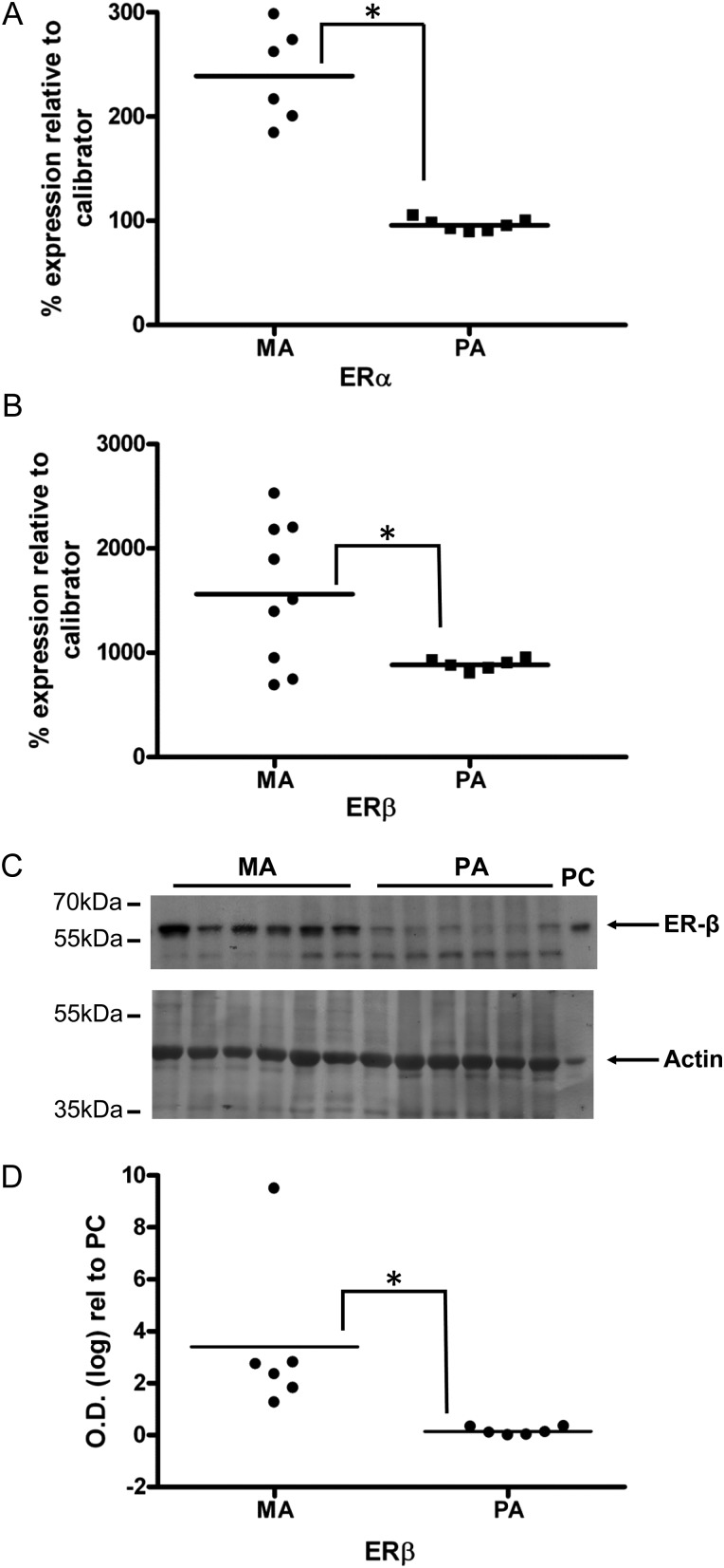
The discrete regulation of vascular tone in the human uterine and placental circulations is a key determinant of appropriate uteroplacental blood perfusion and pregnancy success. Humoral factors such as estrogen, which increases in the placenta and maternal circulation throughout human pregnancy, may regulate these vascular beds as studies of animal arteries have shown that 17β-estradiol, or agonists of estrogen receptors (ER), can exert acute vasodilatory actions. The aim of this study was to compare how acute exposure to ER-specific agonists, and 17β-estradiol, altered human placental and uterine arterial tone in vitro. Uterine and placental arteries were isolated from biopsies obtained from women with uncomplicated pregnancy delivering a singleton infant at term. Vessels were mounted on a wire myograph, exposed to the thromboxane receptor agonist U46619 (10(-6) M), and then incubated with incremental doses (5 min, 0.03-30 µM) of either 17β-estradiol or agonists specific for the ERs ERα (PPT), ERβ (DPN) or the G-protein-coupled estrogen receptor GPER-1 (G1). ERα and ERβ mRNA expression was assessed. 17β-estradiol, PPT and DPN each relaxed myometrial arteries (P < 0.05) in a manner that was partly endothelium-dependent. In contrast, 17β-estradiol or DPN relaxed placental arteries (maximum relaxation to 42 ± 1.1 or 47.6 ± 6.53% of preconstriction, respectively) to a lesser extent than myometrial arteries (to 0.03 ± 0.03 or 8.0 ± 1.0%) and in an endothelial-independent manner whereas PPT was without effect. G1 exposure did not inhibit the constriction of myometrial nor placenta arteries. mRNA expression of ERα and ERβ was greater in myometrial arteries than placental arteries. ER-specific agonists, and 17β-estradiol, differentially modulate the tone of uterine versus placental arteries highlighting that estrogen may regulate human uteroplacental blood flow in a tissue-specific manner.
人类子宫和胎盘循环中血管张力的离散调节是适当的胎盘血流灌注和妊娠成功的关键决定因素。激素等体液因素,如雌激素,在整个妊娠期间在胎盘和母体循环中增加,可能会调节这些血管床,因为动物动脉的研究表明,17β-雌二醇或雌激素受体 (ER) 的激动剂可以发挥急性血管舒张作用。本研究旨在比较 ER 特异性激动剂和 17β-雌二醇急性暴露如何改变体外人胎盘和子宫动脉的张力。从足月分娩单胎婴儿的无并发症妊娠妇女的活检中分离出子宫和胎盘动脉。血管安装在金属丝肌描记器上,暴露于血栓素受体激动剂 U46619(10(-6) M),然后用递增剂量(5 分钟,0.03-30 μM)的 17β-雌二醇或 ER 特异性激动剂孵育,ERα(PPT),ERβ(DPN)或 G 蛋白偶联雌激素受体 GPER-1(G1)。评估 ERα 和 ERβ mRNA 的表达。17β-雌二醇、PPT 和 DPN 均使子宫动脉舒张(P < 0.05),这种舒张部分依赖于内皮。相比之下,17β-雌二醇或 DPN 使胎盘动脉舒张(最大舒张至预收缩的 42 ± 1.1% 或 47.6 ± 6.53%)的程度小于子宫动脉(至 0.03 ± 0.03% 或 8.0 ± 1.0%),并且是非内皮依赖性的,而 PPT 则没有作用。G1 暴露不会抑制子宫和胎盘动脉的收缩。ERα 和 ERβ 的 mRNA 表达在子宫动脉中高于胎盘动脉。ER 特异性激动剂和 17β-雌二醇差异调节子宫和胎盘动脉的张力,这表明雌激素可能以组织特异性方式调节人胎盘血流。