Dogonowski Anne-Marie, Andersen Kasper Winther, Madsen Kristoffer Hougaard, Sørensen Per Soelberg, Paulson Olaf Bjarne, Blinkenberg Morten, Siebner Hartwig Roman
Danish Research Centre for Magnetic Resonance, Centre for Functional and Diagnostic Imaging and Research, Copenhagen University Hospital Hvidovre, Kettegaard Allé 30, 2650 Hvidovre, Denmark.
Danish Research Centre for Magnetic Resonance, Centre for Functional and Diagnostic Imaging and Research, Copenhagen University Hospital Hvidovre, Kettegaard Allé 30, 2650 Hvidovre, Denmark ; Cognitive Systems, Department of Applied Mathematics and Computer Science, Technical University of Denmark, Matematiktorvet, Building 321, 2800 Lyngby, Denmark.
Neuroimage Clin. 2013 Nov 27;4:130-8. doi: 10.1016/j.nicl.2013.11.005. eCollection 2014.
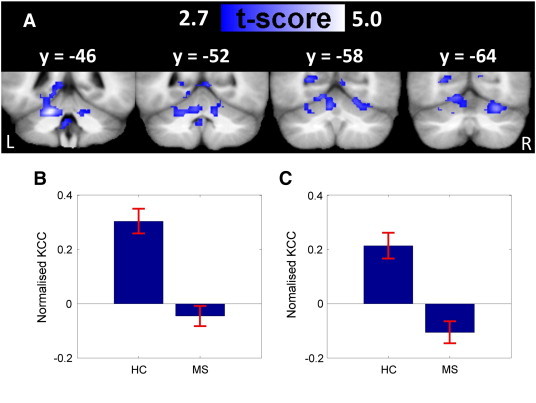
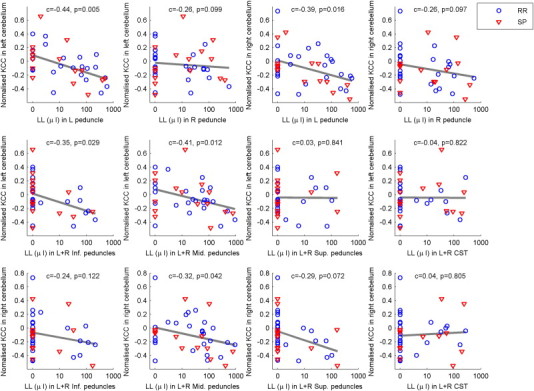
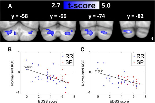
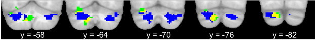
Resting-state functional magnetic resonance imaging (rs-fMRI) has been used to study changes in long-range functional brain connectivity in multiple sclerosis (MS). Yet little is known about how MS affects functional brain connectivity at the local level. Here we studied 42 patients with MS and 30 matched healthy controls with whole-brain rs-fMRI at 3 T to examine local functional connectivity. Using the Kendall's Coefficient of Concordance, regional homogeneity of blood-oxygen-level-dependent (BOLD)-signal fluctuations was calculated for each voxel and used as a measure of local connectivity. Patients with MS showed a decrease in regional homogeneity in the upper left cerebellar hemisphere in lobules V and VI relative to healthy controls. Similar trend changes in regional homogeneity were present in the right cerebellar hemisphere. The results indicate a disintegration of regional processing in the cerebellum in MS. This might be caused by a functional disruption of cortico-ponto-cerebellar and spino-cerebellar inputs, since patients with higher lesion load in the left cerebellar peduncles showed a stronger reduction in cerebellar homogeneity. In patients, two clusters in the left posterior cerebellum expressed a reduction in regional homogeneity with increasing global disability as reflected by the Expanded Disability Status Scale (EDSS) score or higher ataxia scores. The two clusters were mainly located in Crus I and extended into Crus II and the dentate nucleus but with little spatial overlap. These findings suggest a link between impaired regional integration in the cerebellum and general disability and ataxia.
静息态功能磁共振成像(rs-fMRI)已被用于研究多发性硬化症(MS)患者大脑远程功能连接的变化。然而,关于MS如何影响局部水平的大脑功能连接,人们所知甚少。在此,我们对42例MS患者和30名匹配的健康对照者进行了3T全脑rs-fMRI检查,以研究局部功能连接。使用肯德尔和谐系数,计算每个体素的血氧水平依赖(BOLD)信号波动的区域同质性,并将其用作局部连接的指标。与健康对照相比,MS患者左侧小脑半球小叶V和VI的区域同质性降低。右侧小脑半球也出现了类似的区域同质性趋势变化。结果表明MS患者小脑区域处理功能解体。这可能是由于皮质-脑桥-小脑和脊髓-小脑输入的功能破坏所致,因为左侧小脑脚病变负荷较高的患者小脑同质性降低更为明显。在患者中,左侧小脑后部的两个簇随着扩展残疾状态量表(EDSS)评分或更高的共济失调评分所反映的整体残疾程度增加,区域同质性降低。这两个簇主要位于小脑脚I,并延伸至小脑脚II和齿状核,但空间重叠较少。这些发现表明小脑区域整合受损与总体残疾和共济失调之间存在联系。