Mutsaerts Henri J M M, Richard Edo, Heijtel Dennis F R, van Osch Matthias J P, Majoie Charles B L M, Nederveen Aart J
Department of Radiology, Academic Medical Center, Amsterdam, The Netherlands.
Department of Neurology, Academic Medical Center, Amsterdam, The Netherlands.
Neuroimage Clin. 2013 Nov 15;4:139-44. doi: 10.1016/j.nicl.2013.11.003. eCollection 2014.
White matter (WM) perfusion measurements with arterial spin labeling can be severely contaminated by gray matter (GM) perfusion signal, especially in the elderly. The current study investigates the spatial extent of GM contamination by comparing perfusion signal measured in the WM with signal measured outside the brain.
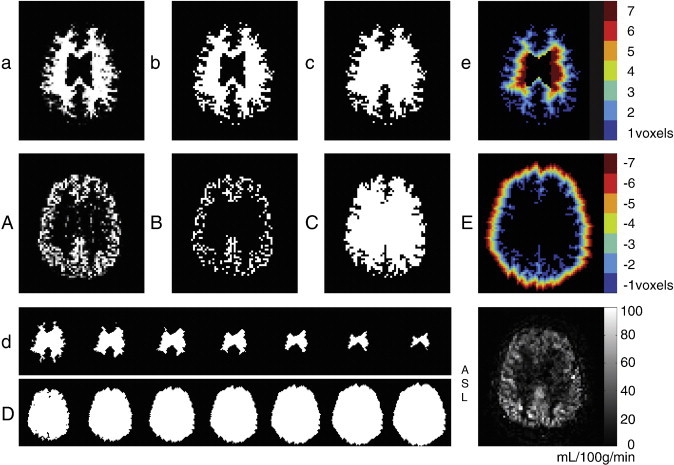
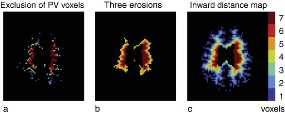
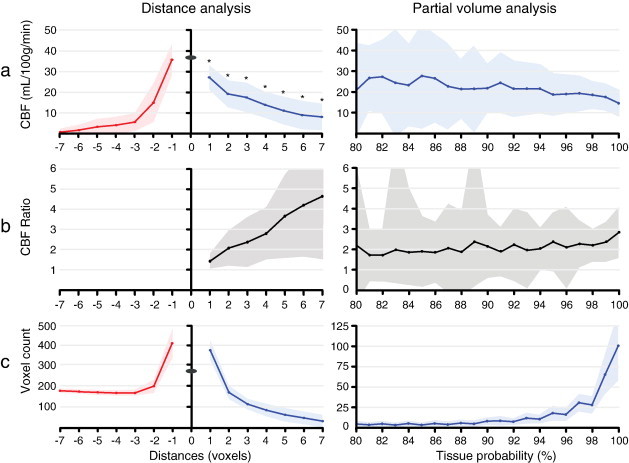
Four minute 3T pseudo-continuous arterial spin labeling scans were performed in 41 elderly subjects with cognitive impairment. Outward and inward geodesic distance maps were created, based on dilations and erosions of GM and WM masks. For all outward and inward geodesic distances, the mean CBF was calculated and compared.
GM contamination was mainly found in the first 3 subcortical WM voxels and had only minor influence on the deep WM signal (distances 4 to 7 voxels). Perfusion signal in the WM was significantly higher than perfusion signal outside the brain, indicating the presence of WM signal.
These findings indicate that WM perfusion signal can be measured unaffected by GM contamination in elderly patients with cognitive impairment. GM contamination can be avoided by the erosion of WM masks, removing subcortical WM voxels from the analysis. These results should be taken into account when exploring the use of WM perfusion as micro-vascular biomarker.
利用动脉自旋标记法测量白质(WM)灌注时,灰质(GM)灌注信号可能会造成严重干扰,在老年人中尤为如此。本研究通过比较脑白质中测量的灌注信号与脑外测量的信号,来探究灰质干扰的空间范围。
对41名患有认知障碍的老年人进行了4分钟的3T伪连续动脉自旋标记扫描。基于灰质和白质掩码的膨胀和腐蚀,创建了向外和向内的测地距离图。计算并比较所有向外和向内测地距离的平均脑血流量(CBF)。
灰质干扰主要出现在皮层下前三组白质体素中,对深部白质信号(距离为4至7个像素)的影响较小。白质中的灌注信号显著高于脑外的灌注信号,表明白质信号的存在。
这些发现表明,在患有认知障碍的老年患者中,可以测量白质灌注信号,且不受灰质干扰的影响。通过对白质掩码进行腐蚀,从分析中去除皮层下白质体素,可以避免灰质干扰。在探索将白质灌注用作微血管生物标志物时,应考虑这些结果。