Promjunyakul N, Lahna D, Kaye J A, Dodge H H, Erten-Lyons D, Rooney W D, Silbert L C
Department of Neurology, Oregon Health & Science University, Portland, OR 97239, USA.
Department of Neurology, Oregon Health & Science University, Portland, OR 97239, USA ; Department of Neurology, Veterans Affairs Medical Center, Portland, OR 97239, USA.
Neuroimage Clin. 2015 Apr 22;8:224-9. doi: 10.1016/j.nicl.2015.04.012. eCollection 2015.
White matter hyperintensities (WMHs) are common with age, grow over time, and are associated with cognitive and motor impairments. Mechanisms underlying WMH growth are unclear. We aimed to determine the presence and extent of decreased normal appearing white matter (NAWM) cerebral blood flow (CBF) surrounding WMHs to identify 'WM at risk', or the WMH CBF penumbra. We aimed to further validate cross-sectional finding by determining whether the baseline WMH penumbra CBF predicts the development of new WMHs at follow-up.
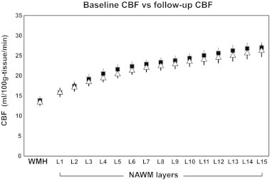
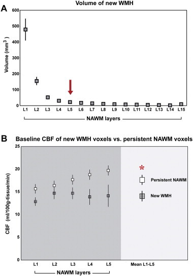
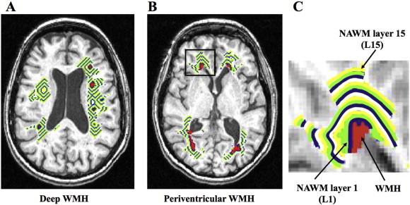
Sixty-one cognitively intact elderly subjects received 3 T MPRAGE, FLAIR, and pulsed arterial spin labeling (PASL). Twenty-four subjects returned for follow-up MRI. The inter-scan interval was 18 months. A NAWM layer mask, comprised of fifteen layers, 1 mm thick each surrounding WMHs, was generated for periventricular (PVWMH) and deep (DWMH) WMHs. Mean CBF for each layer was computed. New WMH and persistent NAWM voxels for each penumbra layer were defined from follow-up MRI.
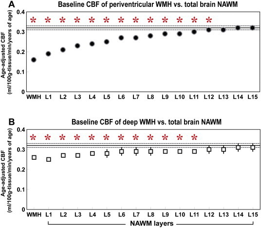
CBF in the area surrounding WMHs was significantly lower than the total brain NAWM, extending approximately 12 mm from both the established PVWMH and DWMH. Voxels with new WMH at follow-up had significantly lower baseline CBF than voxels that maintained NAWM, suggesting that baseline CBF can predict the development of new WMHs over time.
A CBF penumbra exists surrounding WMHs, which is associated with future WMH expansion. ASL MRI can be used to monitor interventions to increase white matter blood flow for the prevention of further WM damage and its cognitive and motor consequences.
脑白质高信号(WMHs)随年龄增长而常见,且会随时间增加,并与认知和运动障碍相关。WMHs增加的潜在机制尚不清楚。我们旨在确定WMHs周围正常脑白质(NAWM)脑血流量(CBF)降低的存在情况及范围,以识别“有风险的白质”或WMH CBF半暗带。我们旨在通过确定基线WMH半暗带CBF是否能预测随访中新WMHs的发生,进一步验证横断面研究结果。
61名认知功能正常的老年人接受了3T的MPRAGE、FLAIR和脉冲动脉自旋标记(PASL)检查。24名受试者返回进行随访MRI检查。扫描间隔为18个月。为脑室周围(PVWMH)和深部(DWMH)WMHs生成了一个由15层组成的NAWM层掩码,每层厚度为1mm,围绕WMHs。计算每层的平均CBF。从随访MRI中定义每个半暗带层的新WMH和持续的NAWM体素。
WMHs周围区域的CBF显著低于全脑NAWM,从已确定的PVWMH和DWMH均向外延伸约12mm。随访时出现新WMH的体素的基线CBF显著低于维持NAWM的体素,这表明基线CBF可预测随时间推移新WMHs的发生。
WMHs周围存在CBF半暗带,这与未来WMH的扩大相关。动脉自旋标记MRI可用于监测增加白质血流量的干预措施,以预防进一步的白质损伤及其认知和运动后果。