Laboratory of Cellular and Molecular Neurobiology, Graduate School of Frontier Biosciences, Osaka University, Suita, Osaka, Japan.
Department of Molecular Neuroscience, Graduate School of Medicine, Osaka University, Suita, Osaka, Japan.
PLoS One. 2013 Dec 23;8(12):e82954. doi: 10.1371/journal.pone.0082954. eCollection 2013.
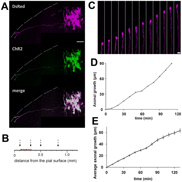
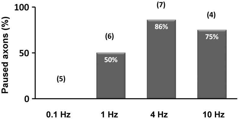
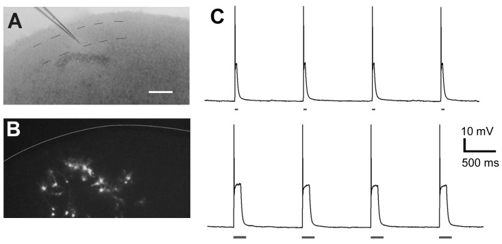
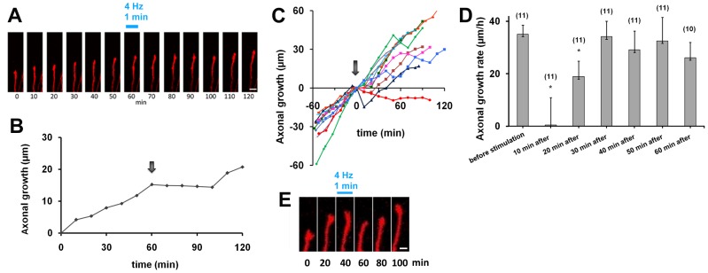
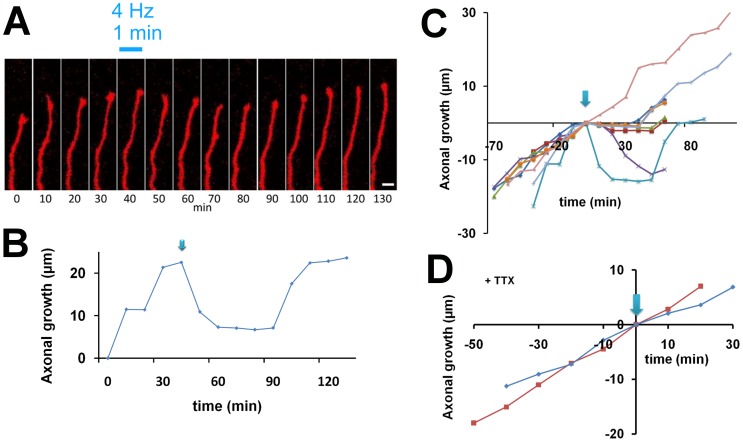
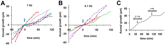
During development, layer 2/3 neurons in the neocortex extend their axons horizontally, within the same layers, and stop growing at appropriate locations to form branches and synaptic connections. Firing and synaptic activity are thought to be involved in this process, but how neuronal activity regulates axonal growth is not clear. Here, we studied axonal growth of layer 2/3 neurons by exciting cell bodies or axonal processes in organotypic slice cultures of the rat cortex. For neuronal stimulation and morphological observation, plasmids encoding channelrhodopsin-2 (ChR2) and DsRed were coelectroporated into a small number of layer 2/3 cells. Firing activity induced by photostimulation (475 nm) was confirmed by whole-cell patch recording. Axonal growth was observed by time-lapse confocal microscopy, using a different excitation wavelength (560 nm), at 10-20-min intervals for several hours. During the first week in vitro, when spontaneous neuronal activity is low, DsRed- and ChR2-expressing axons grew at a constant rate. When high-frequency photostimulation (4 or 10 Hz) for 1 min was applied to the soma or axon, most axons paused in their growth. In contrast, lower-frequency stimulation did not elicit this pause behavior. Moreover, in the presence of tetrodotoxin, even high-frequency stimulation did not cause axonal growth to pause. These results indicate that increasing firing activity during development suppresses axon growth, suggesting the importance of neuronal activity for the formation of horizontal connections.
在发育过程中,皮质的 2/3 层神经元的轴突在同一层内水平延伸,并在适当的位置停止生长,形成分支和突触连接。据认为,放电和突触活动参与了这一过程,但神经元活动如何调节轴突生长尚不清楚。在这里,我们通过在大鼠皮质的器官型切片培养物中兴奋细胞体或轴突过程来研究 2/3 层神经元的轴突生长。为了进行神经元刺激和形态观察,将编码通道视紫红质-2(ChR2)和 DsRed 的质粒共转染到少量 2/3 层细胞中。通过全细胞膜片钳记录确认光刺激(475nm)引起的放电活动。通过时间 lapse 共聚焦显微镜,使用不同的激发波长(560nm),每隔 10-20 分钟观察数小时,观察轴突生长。在体外培养的第一周,当自发神经元活动较低时,DsRed 和 ChR2 表达的轴突以恒定的速度生长。当对细胞体或轴突进行 1 分钟的高频光刺激(4 或 10Hz)时,大多数轴突在生长中暂停。相比之下,低频刺激不会引起这种暂停行为。此外,在加入河豚毒素的情况下,即使是高频刺激也不会导致轴突生长暂停。这些结果表明,发育过程中放电活动的增加抑制了轴突生长,这表明神经元活动对于形成水平连接的重要性。