Ni Na, Hu Yaohua, Ren Huixia, Luo Chuanming, Li Peng, Wan Jian-Bo, Su Huanxing
State Key Laboratory of Quality Research in Chinese Medicine, Institute of Chinese Medical Sciences, University of Macau, Macao, China.
PLoS One. 2013 Dec 20;8(12):e84504. doi: 10.1371/journal.pone.0084504. eCollection 2013.
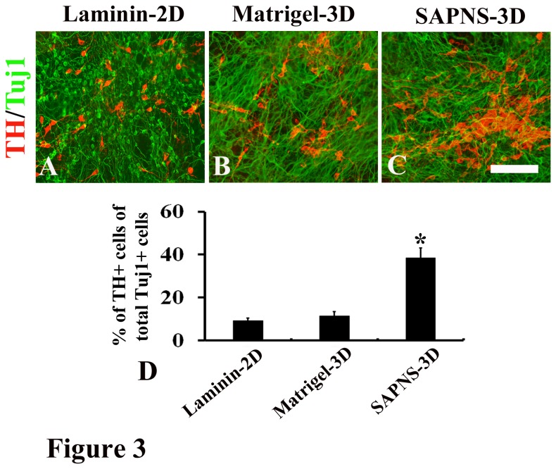
Dopaminergic differentiation of embryonic stem cells (ESCs) gains more and more attention worldwide owing to its potential use for neurorestorative therapy for the treatment of Parkinson's disease. The conventional 2D cell culture on petri dishes with various animal derived substrata such as collagen gels, laminin, and Matrigel is widely used to induce dopaminergic differentiation and it may limit the efficiency in the generation of dopaminergic neurons from ESCs and prevent their application for human therapies. Here, we reported that a self-assembling peptide made from natural amino acids has a property to generate a true 3D environment for dopaminergic differentiation. Mouse ESCs (R1) and mouse iPSCs (TTF-1) embedded in RADA16-I peptide-derived nanofiber scaffolds led to a marked increase in dopaminergic differentiation compared to the laminin-coated 2D culture or Matrigel-encapsulated 3D culture. These differentiated neurons expressed specific dopaminergic markers and produced appropriate patterns of action potential firing. Consistent with the increase in the number of dopaminergic neurons differentiated from R1 or TTF-1 in the self-assembling peptide nanofiber scaffold (SAPNS), both the expression levels of genes that involve in dopaminergic differentiation and maturation and the dopamine release in SAPNS culture were significantly elevated. The results of the study suggest that SAPNS provides a promising 3D culture system for dopaminergic differentiation.
胚胎干细胞(ESC)的多巴胺能分化因其在帕金森病神经修复治疗中的潜在应用而在全球范围内受到越来越多的关注。传统的在培养皿上使用各种动物源性基质(如胶原蛋白凝胶、层粘连蛋白和基质胶)进行的二维细胞培养被广泛用于诱导多巴胺能分化,但这可能会限制从胚胎干细胞中产生多巴胺能神经元的效率,并阻碍其在人类治疗中的应用。在此,我们报道了一种由天然氨基酸制成的自组装肽具有为多巴胺能分化生成真实三维环境的特性。与层粘连蛋白包被的二维培养或基质胶包封的三维培养相比,嵌入RADA16-I肽衍生的纳米纤维支架中的小鼠胚胎干细胞(R1)和小鼠诱导多能干细胞(TTF-1)导致多巴胺能分化显著增加。这些分化的神经元表达特定的多巴胺能标记物,并产生适当的动作电位发放模式。与在自组装肽纳米纤维支架(SAPNS)中从R1或TTF-1分化出的多巴胺能神经元数量增加一致,参与多巴胺能分化和成熟的基因表达水平以及SAPNS培养中的多巴胺释放均显著升高。该研究结果表明,SAPNS为多巴胺能分化提供了一个有前景的三维培养系统。