Akkina R K, Chambers T M, Londo D R, Nayak D P
J Virol. 1987 Jul;61(7):2217-24. doi: 10.1128/JVI.61.7.2217-2224.1987.
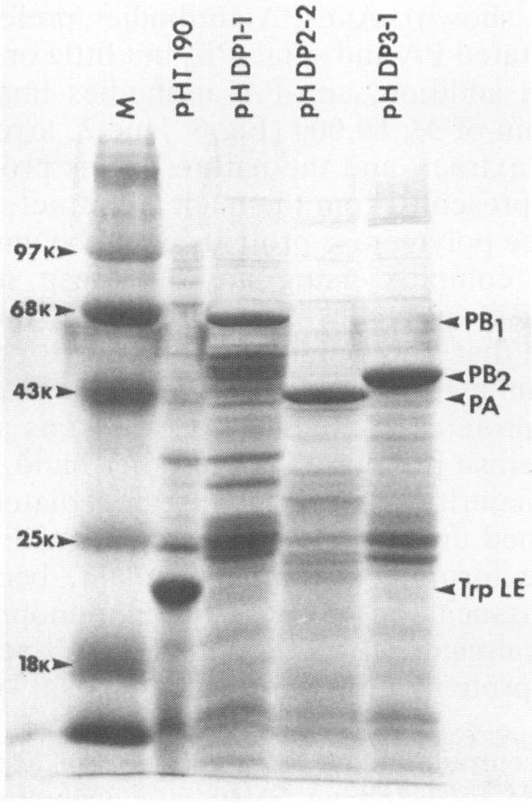
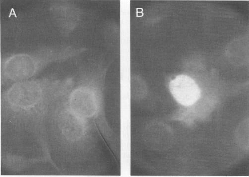
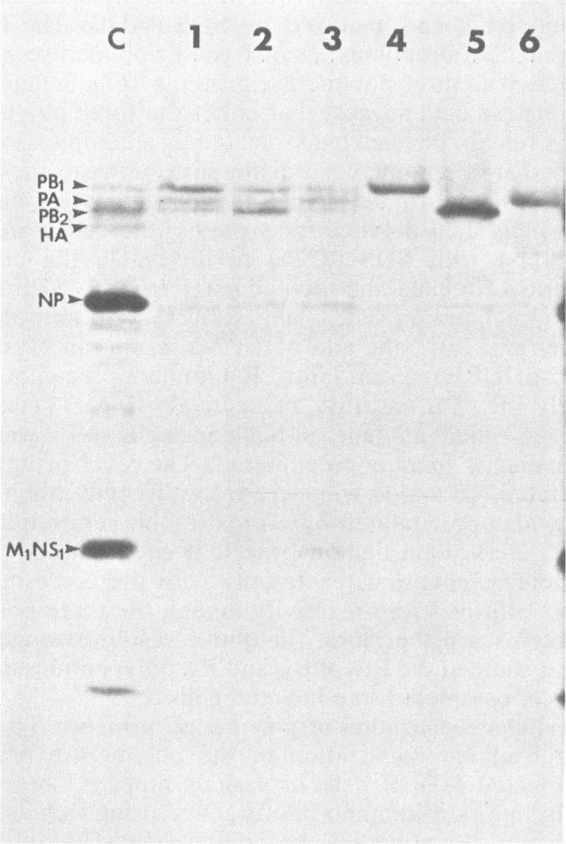
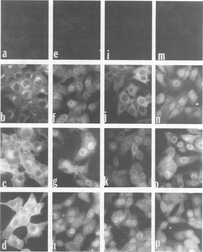
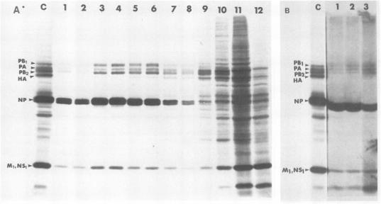
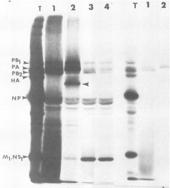
The biosynthesis, nuclear transport, and formation of a complex among the influenza polymerase proteins were studied in influenza virus-infected MDBK cells by using monospecific antisera. To obtain these monospecific antisera, portions of cloned cDNAs encoding the individual polymerase proteins (PB1, PB2, or PA) of A/WSN/33 influenza virus were expressed as fusion proteins in Escherichia coli, and the purified fusion proteins were injected into rabbits. Studies using indirect immunofluorescence showed that early in the infectious cycle (4 h postinfection) of influenza virus, PB1 and PB2 are present mainly in the nucleus, whereas PA is predominantly present in the cytoplasm of the virus-infected cells. Later, at 6 to 8 h postinfection, all three polymerase proteins are apparent both in the cytoplasm as well as the nucleus. Radiolabeling and immunoprecipitation analyses showed that the three polymerase proteins remain physically associated as a complex in either the presence or the absence of ribonucleoproteins. In the cytoplasm, the majority of the polymerase proteins remain unassociated, whereas in the nucleus they are present as a complex of three polymerase proteins. To determine whether a polymerase protein is transported into the nucleus individually, PB1 was expressed from the cloned cDNA by using the simian virus 40 late promoter expression vector. PB1 alone, in the absence of the other polymerase proteins or the nucleoprotein, accumulates in the nucleus. This suggests that the formation of a complex with other viral protein(s) is not required for either nuclear transport or nuclear accumulation of PB1 protein and that the PB1 protein may contain an intrinsic signal(s) for nuclear transport.
利用单特异性抗血清,在感染流感病毒的MDBK细胞中研究了流感病毒聚合酶蛋白的生物合成、核转运及复合物形成。为获得这些单特异性抗血清,将编码A/WSN/33流感病毒单个聚合酶蛋白(PB1、PB2或PA)的部分克隆cDNA在大肠杆菌中表达为融合蛋白,然后将纯化的融合蛋白注射到兔子体内。间接免疫荧光研究表明,在流感病毒感染周期早期(感染后4小时),PB1和PB2主要存在于细胞核中,而PA主要存在于病毒感染细胞的细胞质中。之后,在感染后6至8小时,所有三种聚合酶蛋白在细胞质和细胞核中均可见。放射性标记和免疫沉淀分析表明,无论有无核糖核蛋白,这三种聚合酶蛋白均以复合物形式保持物理结合。在细胞质中,大多数聚合酶蛋白未结合,而在细胞核中它们以三种聚合酶蛋白的复合物形式存在。为确定聚合酶蛋白是否单独转运至细胞核,利用猿猴病毒40晚期启动子表达载体从克隆的cDNA中表达PB1。单独的PB1在没有其他聚合酶蛋白或核蛋白的情况下积聚在细胞核中。这表明PB1蛋白的核转运或核积聚不需要与其他病毒蛋白形成复合物,并且PB1蛋白可能含有核转运的内在信号。