Department of Disaster Medicine, Lithuanian University of Health Sciences, Eiveniu 4, LT-50161 Kaunas, Lithuania.
BMC Neurosci. 2014 Jan 3;15:2. doi: 10.1186/1471-2202-15-2.
Ischemic brain injury due to stroke and/or cardiac arrest is a major health issue in modern society requiring urgent development of new effective therapies. The aim of this study was to evaluate mitochondrial, microcirculatory, and histological changes in a swine model of global cerebral ischemia.
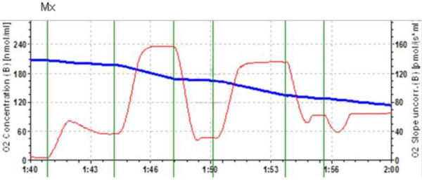
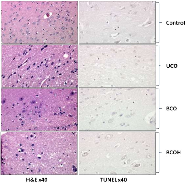
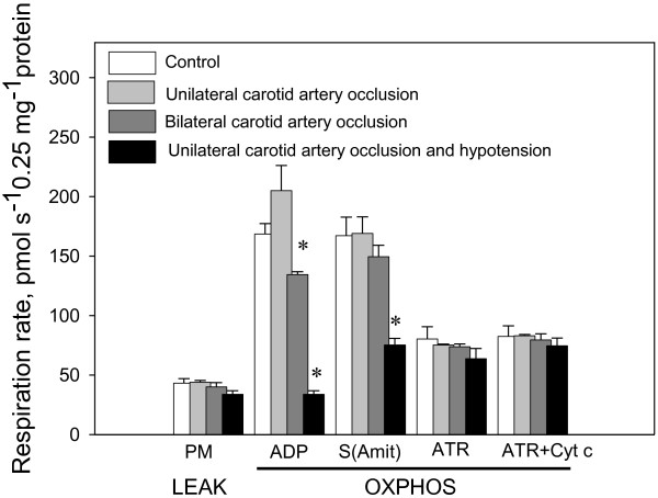
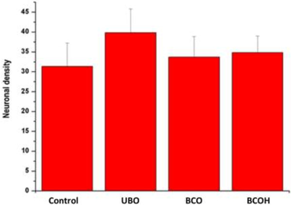
In our model, significant microcirculatory changes, but only negligible histological cell alterations, were observed 3 h after bilateral carotid occlusion, and were more pronounced if the vascular occlusion was combined with systemic hypotension. Analysis of mitochondrial function showed that LEAK respiration (measured in the presence of pyruvate + malate but without ADP) was not affected in any model of global cerebral ischemia in pigs. The OXPHOS capacity with pyruvate + malate as substrates decreased compared with the control levels after bilateral carotid artery occlusion, and bilateral carotid artery occlusion + hypotension by 20% and 79%, respectively, resulting in decreases in the respiratory control index of 14% and 73%, respectively. OXPHOS capacity with succinate as a substrate remained constant after unilateral carotid artery occlusion or bilateral carotid artery occlusion, but decreased by 53% after bilateral carotid artery occlusion and hypotension compared with controls (p < 0.05, n = 3-6). Addition of exogenous cytochrome c to mitochondria isolated from ischemia brains had no effect on respiration in all models used in this study.
We found a decrease in microcirculation and mitochondrial oxidative phosphorylation activity, but insignificant neuronal death, after 3 h ischemia in all our pig models of global cerebral ischemia. Dysfunction of the mitochondrial oxidative phosphorylation system, particularly damage to complex I of the respiratory chain, may be the primary target of the ischemic insult, and occurs before signs of neuronal death can be detected.
由于中风和/或心脏骤停导致的缺血性脑损伤是现代社会的一个主要健康问题,需要迫切开发新的有效治疗方法。本研究旨在评估猪全脑缺血模型中的线粒体、微循环和组织学变化。
在我们的模型中,双侧颈总动脉闭塞 3 小时后观察到明显的微循环变化,但只有可忽略的组织学细胞改变,如果血管闭塞与全身低血压相结合,则更为明显。线粒体功能分析表明,在存在丙酮酸+苹果酸但没有 ADP 的情况下测量的 LEAK 呼吸(泄漏呼吸)在任何猪全脑缺血模型中均不受影响。与对照组相比,丙酮酸+苹果酸作为底物的 OXPHOS 能力在双侧颈总动脉闭塞后下降,双侧颈总动脉闭塞+低血压分别下降 20%和 79%,导致呼吸控制指数分别下降 14%和 73%。单侧颈总动脉闭塞或双侧颈总动脉闭塞后,以琥珀酸作为底物的 OXPHOS 能力保持不变,但与对照组相比,双侧颈总动脉闭塞和低血压后下降 53%(p<0.05,n=3-6)。在本研究中使用的所有模型中,将外源性细胞色素 c 添加到从缺血大脑中分离的线粒体中对呼吸没有影响。
我们发现,在所有猪全脑缺血模型中,缺血 3 小时后微循环和线粒体氧化磷酸化活性下降,但神经元死亡不明显。线粒体氧化磷酸化系统功能障碍,特别是呼吸链复合物 I 的损伤,可能是缺血损伤的主要靶点,并且发生在可检测到神经元死亡之前。