• 文献检索
  • 文档翻译
  • 深度研究
  • 学术资讯
  • Suppr Zotero 插件Zotero 插件
  • 邀请有礼
  • 套餐&价格
  • 历史记录
应用&插件
Suppr Zotero 插件Zotero 插件浏览器插件Mac 客户端Windows 客户端微信小程序
定价
高级版会员购买积分包购买API积分包
服务
文献检索文档翻译深度研究API 文档MCP 服务
关于我们
关于 Suppr公司介绍联系我们用户协议隐私条款
关注我们

Suppr 超能文献

核心技术专利:CN118964589B侵权必究
粤ICP备2023148730 号-1Suppr @ 2026

文献检索

告别复杂PubMed语法,用中文像聊天一样搜索,搜遍4000万医学文献。AI智能推荐,让科研检索更轻松。

立即免费搜索

文件翻译

保留排版,准确专业,支持PDF/Word/PPT等文件格式,支持 12+语言互译。

免费翻译文档

深度研究

AI帮你快速写综述,25分钟生成高质量综述,智能提取关键信息,辅助科研写作。

立即免费体验

囊袋警告综合征患者的高分辨率 MRI 表现。

High-resolution MRI findings in patients with capsular warning syndrome.

机构信息

Department of Neurology, Peking Union Medical College Hospital and Chinese Academy of Medical Science, Shuai Fu Yuan 1#, Dong Cheng District, Beijing 100730, China.

出版信息

BMC Neurol. 2014 Jan 20;14:16. doi: 10.1186/1471-2377-14-16.

DOI:10.1186/1471-2377-14-16
PMID:24438445
原文链接:https://pmc.ncbi.nlm.nih.gov/articles/PMC3898091/
Abstract

BACKGROUND

Capsular warning syndrome (CWS) is rare (1.5% of TIA presentations) but has a poor prognosis (7-day stroke risk of 60%). Up to date, the exact pathogenic mechanism of CWS has not been fully understood. We report the clinical presentations and high-resolution MRI (HR MRI) findings of two cases with capsular warning symptoms.

CASE PRESENTATION

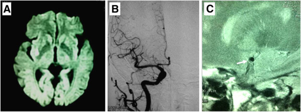
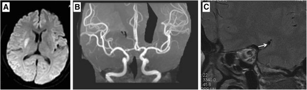
Case 1 was a 63-year-old man with a history of hypertension with recurrent episodes of left hemiparesis and dysarthria lasting 10 ~ 30 minutes. Case 2 was a 54-year-old woman with repetitive episodes of transient left hemiparesis and dysarthria lasting about 10 minutes. Capsular infarctions on DWI were demonstrated in the territory of a lenticulostriate artery in both 2 patients. HR MRI disclosed atherosclerotic plaques on the ventral wall of the MCA where enticulostriate arteries were arisen from, although traditional digital subtraction angiography showed normal. Aggressive medical therapy with dual antithrombotic agents and statin was effective in these two cases.

CONCLUSION

Our HR MRI data offer an insight into the pathophysiology of CWS which might be caused by atherosclerotic plaque in non-stenotic MCA wall. HR MRI might be a useful modality for characterizing atherosclerotic plaques in the MCA and detecting the pathophysiology of the CWS.

摘要

背景

囊袋警告综合征(CWS)较为罕见(占 TIA 表现的 1.5%),但其预后较差(7 天内卒中风险为 60%)。迄今为止,CWS 的确切发病机制尚未完全阐明。我们报告了两例具有囊袋警告症状的病例的临床表现和高分辨率 MRI(HR MRI)结果。

病例介绍

病例 1 为 63 岁男性,有高血压病史,反复出现左侧偏瘫和构音障碍,持续 10~30 分钟。病例 2 为 54 岁女性,反复出现短暂性左侧偏瘫和构音障碍,持续约 10 分钟。两例患者 DWI 均显示壳核梗死,位于纹状体动脉供血区。尽管传统的数字减影血管造影显示正常,但 HR MRI 显示 MCA 腹侧壁存在粥样硬化斑块,纹状体动脉由此发出。这两例患者均采用双联抗血栓药物和他汀类药物进行强化治疗,效果良好。

结论

我们的 HR MRI 数据提供了对 CWS 病理生理学的深入了解,其可能是由非狭窄性 MCA 壁中的粥样硬化斑块引起的。HR MRI 可能是一种用于表征 MCA 中粥样硬化斑块和检测 CWS 病理生理学的有用方法。

https://cdn.ncbi.nlm.nih.gov/pmc/blobs/6077/3898091/273f40a98d1b/1471-2377-14-16-2.jpg
https://cdn.ncbi.nlm.nih.gov/pmc/blobs/6077/3898091/43b6d4c0e018/1471-2377-14-16-1.jpg
https://cdn.ncbi.nlm.nih.gov/pmc/blobs/6077/3898091/273f40a98d1b/1471-2377-14-16-2.jpg
https://cdn.ncbi.nlm.nih.gov/pmc/blobs/6077/3898091/43b6d4c0e018/1471-2377-14-16-1.jpg
https://cdn.ncbi.nlm.nih.gov/pmc/blobs/6077/3898091/273f40a98d1b/1471-2377-14-16-2.jpg

相似文献

1
High-resolution MRI findings in patients with capsular warning syndrome.囊袋警告综合征患者的高分辨率 MRI 表现。
BMC Neurol. 2014 Jan 20;14:16. doi: 10.1186/1471-2377-14-16.
2
Value of higher-resolution MRI in assessing middle cerebral atherosclerosis and predicting capsular warning syndrome.高分辨率MRI在评估大脑中动脉粥样硬化及预测壳核预警综合征中的价值
J Magn Reson Imaging. 2016 Nov;44(5):1277-1283. doi: 10.1002/jmri.25265. Epub 2016 Apr 15.
3
Capsular warning syndrome caused by middle cerebral artery stenosis.大脑中动脉狭窄致囊袋警告综合征。
J Neurol Sci. 2010 Sep 15;296(1-2):115-20. doi: 10.1016/j.jns.2010.06.003. Epub 2010 Jul 8.
4
Loading dose of clopidogrel in combination with other antithrombotic therapy for capsular warning syndrome.氯吡格雷负荷剂量联合其他抗血栓治疗用于囊袋警告综合征。
J Stroke Cerebrovasc Dis. 2014 May-Jun;23(5):1265-6. doi: 10.1016/j.jstrokecerebrovasdis.2013.09.020. Epub 2013 Oct 15.
5
rt-PA with Antithrombotic Therapies in a Case with Capsular Warning Syndrome.重组组织型纤溶酶原激活剂联合抗栓治疗用于一例伴有包膜警告综合征的病例
Intern Med. 2017;56(4):441-444. doi: 10.2169/internalmedicine.56.7522. Epub 2017 Feb 15.
6
Capsular warning syndrome and its clinical awareness and therapeutic approach: two case reports and a systematic review of the literature.包膜警告综合征及其临床认识与治疗方法:两例病例报告及文献系统综述
Front Neurol. 2023 May 16;14:1177660. doi: 10.3389/fneur.2023.1177660. eCollection 2023.
7
High resolution magnetic resonance imaging in pathogenesis diagnosis of single lenticulostriate infarction with nonstenotic middle cerebral artery, a retrospective study.高分辨率磁共振成像在非狭窄性大脑中动脉所致单纯豆纹状核梗死发病机制诊断中的应用:一项回顾性研究
BMC Neurol. 2018 Apr 25;18(1):51. doi: 10.1186/s12883-018-1054-z.
8
Diffusion-weighted MRI findings in patients with capsular warning syndrome.包膜警告综合征患者的扩散加权磁共振成像结果
Cerebrovasc Dis. 2004;17(1):1-8. doi: 10.1159/000073891. Epub 2003 Oct 3.
9
Cerebral Perforating Artery Disease : Characteristics on High-Resolution Magnetic Resonance Imaging.穿支动脉疾病:高分辨率磁共振成像特征。
Clin Neuroradiol. 2019 Sep;29(3):533-541. doi: 10.1007/s00062-018-0682-4. Epub 2018 Mar 23.
10
Capsular warning syndrome-a case of atrial fibrillation and corona radiata infarct.包膜警告综合征——一例心房颤动与放射冠梗死病例
Int J Emerg Med. 2023 Sep 26;16(1):62. doi: 10.1186/s12245-023-00541-w.

引用本文的文献

1
Capsular warning syndrome and its clinical awareness and therapeutic approach: two case reports and a systematic review of the literature.包膜警告综合征及其临床认识与治疗方法:两例病例报告及文献系统综述
Front Neurol. 2023 May 16;14:1177660. doi: 10.3389/fneur.2023.1177660. eCollection 2023.
2
Intravenous tirofiban therapy for patients with capsular warning syndrome.静脉注射替罗非班治疗囊袋警告综合征患者。
Stroke Vasc Neurol. 2019 Jan 9;4(1):22-27. doi: 10.1136/svn-2018-000163. eCollection 2019 Mar.
3
Vessel wall imaging for intracranial vascular disease evaluation.

本文引用的文献

1
Population-based study of capsular warning syndrome and prognosis after early recurrent TIA.基于人群的胶囊警告综合征研究及早期复发性 TIA 后的预后。
Neurology. 2012 Sep 25;79(13):1356-62. doi: 10.1212/WNL.0b013e31826c1af8. Epub 2012 Sep 12.
2
Plaque distribution of stenotic middle cerebral artery and its clinical relevance.狭窄大脑中动脉的斑块分布及其临床意义。
Stroke. 2011 Oct;42(10):2957-9. doi: 10.1161/STROKEAHA.111.618132. Epub 2011 Jul 28.
3
In vivo high-resolution MR imaging of symptomatic and asymptomatic middle cerebral artery atherosclerotic stenosis.
用于颅内血管疾病评估的血管壁成像。
J Neurointerv Surg. 2016 Nov;8(11):1154-1159. doi: 10.1136/neurintsurg-2015-012127. Epub 2016 Jan 14.
4
High-resolution intracranial vessel wall imaging: imaging beyond the lumen.高分辨率颅内血管壁成像:超越管腔的成像
J Neurol Neurosurg Psychiatry. 2016 Jun;87(6):589-97. doi: 10.1136/jnnp-2015-312020. Epub 2016 Jan 8.
5
Nonstenotic Culprit Plaque: The Utility of High-Resolution Vessel Wall MRI of Intracranial Vessels after Ischemic Stroke.无狭窄责任斑块:缺血性卒中后颅内血管高分辨率血管壁磁共振成像的效用
Case Rep Radiol. 2015;2015:356582. doi: 10.1155/2015/356582. Epub 2015 Aug 6.
症状性和无症状性大脑中动脉粥样硬化性狭窄的活体高分辨率磁共振成像。
Atherosclerosis. 2010 Oct;212(2):507-11. doi: 10.1016/j.atherosclerosis.2010.06.035. Epub 2010 Jun 25.
4
Capsular warning syndrome caused by middle cerebral artery stenosis.大脑中动脉狭窄致囊袋警告综合征。
J Neurol Sci. 2010 Sep 15;296(1-2):115-20. doi: 10.1016/j.jns.2010.06.003. Epub 2010 Jul 8.
5
Atherosclerosis of middle cerebral artery: evaluation with high-resolution MR imaging at 3T.大脑中动脉粥样硬化:3T高分辨率磁共振成像评估
Atherosclerosis. 2009 Jun;204(2):447-52. doi: 10.1016/j.atherosclerosis.2008.10.019. Epub 2008 Oct 25.
6
Pontine warning syndrome.脑桥预警综合征
Arch Neurol. 2008 Oct;65(10):1375-7. doi: 10.1001/archneur.65.10.1375.
7
Middle cerebral artery plaque imaging using 3-Tesla high-resolution MRI.使用3特斯拉高分辨率磁共振成像对大脑中动脉斑块进行成像。
J Clin Neurosci. 2008 Oct;15(10):1137-41. doi: 10.1016/j.jocn.2007.09.024. Epub 2008 Aug 13.
8
Thrombolysis in capsular warning syndrome.囊状预警综合征中的溶栓治疗。
Cerebrovasc Dis. 2008;25(5):508-10. doi: 10.1159/000131109. Epub 2008 May 9.
9
In vivo middle cerebral artery plaque imaging by high-resolution MRI.通过高分辨率磁共振成像进行大脑中动脉斑块的体内成像。
Neurology. 2006 Jul 25;67(2):327-9. doi: 10.1212/01.wnl.0000225074.47396.71.
10
Oral clopidogrel load in aspirin-resistant capsular warning syndrome.阿司匹林抵抗性包膜警告综合征中的口服氯吡格雷负荷量
Neurocrit Care. 2005;2(2):183-4. doi: 10.1385/NCC:2:2:183.