Computational Biomedicine Laboratory, Rochester Institute of Technology, Rochester, NY 14623, USA.
Department of Pharmacology, SUNY Upstate Medical University, Syracuse, NY 13210, USA.
Comput Math Methods Med. 2013;2013:293069. doi: 10.1155/2013/293069. Epub 2013 Dec 23.
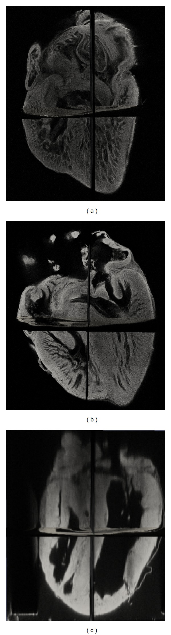
Embryonic heart morphogenesis (EHM) is a complex and dynamic process where the heart transforms from a single tube into a four-chambered pump. This process is of great biological and clinical interest but is still poorly understood for two main reasons. On the one hand, the existing imaging modalities for investigating EHM suffered from either limited penetration depth or limited spatial resolution. On the other hand, current works typically adopted manual segmentation, which was tedious, subjective, and time consuming considering the complexity of developing heart geometry and the large size of images. In this paper, we propose to utilize confocal microscopy imaging with tissue optical immersion clearing technique to image the heart at different stages of development for EHM study. The imaging method is able to produce high spatial resolution images and achieve large penetration depth at the same time. Furthermore, we propose a novel convex active contour model for automatic image segmentation. The model has the ability to deal with intensity fall-off in depth which is characterized by confocal microscopy images. We acquired the images of embryonic quail hearts from day 6 to day 14 of incubation for EHM study. The experimental results were promising and provided us with an insight view of early heart growth pattern and also paved the road for data-driven heart growth modeling.
胚胎心脏形态发生 (EHM) 是一个复杂而动态的过程,在此过程中,心脏从单个管腔转变为具有四个腔室的泵。这一过程具有重要的生物学和临床意义,但由于两个主要原因,其仍然知之甚少。一方面,用于研究 EHM 的现有成像方式要么穿透深度有限,要么空间分辨率有限。另一方面,目前的工作通常采用手动分割,考虑到心脏发育的复杂性和图像的尺寸较大,这种方法既繁琐又耗时,而且具有主观性。在本文中,我们提出利用共聚焦显微镜成像和组织光学浸液清除技术对心脏进行成像,以研究 EHM。该成像方法能够同时获得高空间分辨率的图像和大穿透深度。此外,我们还提出了一种新的凸主动轮廓模型用于自动图像分割。该模型能够处理共聚焦显微镜图像的特征强度衰减。我们采集了孵化第 6 天至第 14 天的鸡胚心脏图像用于 EHM 研究。实验结果令人鼓舞,为我们提供了早期心脏生长模式的深入了解,也为基于数据的心脏生长建模铺平了道路。