Turóczi Zsolt, Arányi Péter, Lukáts Ákos, Garbaisz Dávid, Lotz Gábor, Harsányi László, Szijártó Attila
1st Department of Surgery, Semmelweis University, Budapest, Hungary.
Department of Human Morphology and Developmental Biology, Semmelweis University, Budapest, Hungary.
PLoS One. 2014 Jan 13;9(1):e84783. doi: 10.1371/journal.pone.0084783. eCollection 2014.
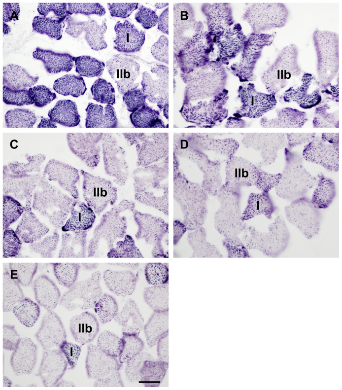
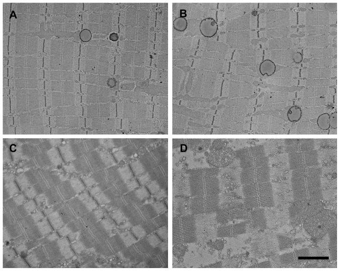
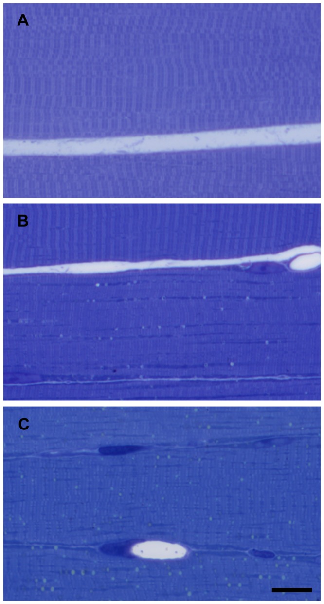
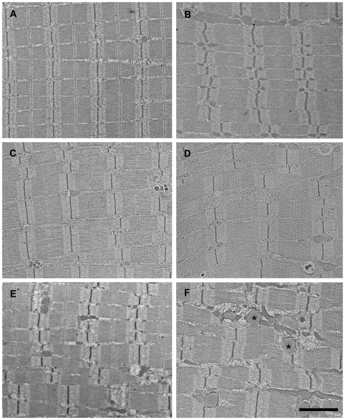
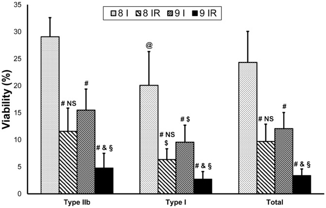
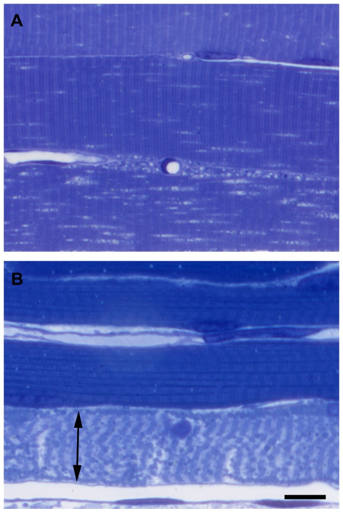
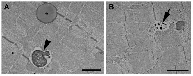
Acute lower extremity ischemia is a limb- and life-threatening clinical problem. Rapid detection of the degree of injury is crucial, however at present there are no exact diagnostic tests available to achieve this purpose. Our goal was to examine a novel technique - which has the potential to accurately assess the degree of ischemic muscle injury within a short period of time - in a clinically relevant rodent model. Male Wistar rats were exposed to 4, 6, 8 and 9 hours of bilateral lower limb ischemia induced by the occlusion of the infrarenal aorta. Additional animals underwent 8 and 9 hours of ischemia followed by 2 hours of reperfusion to examine the effects of revascularization. Muscle samples were collected from the left anterior tibial muscle for viability assessment. The degree of muscle damage (muscle fiber viability) was assessed by morphometric evaluation of NADH-tetrazolium reductase reaction on frozen sections. Right hind limbs were perfusion-fixed with paraformaldehyde and glutaraldehyde for light and electron microscopic examinations. Muscle fiber viability decreased progressively over the time of ischemia, with significant differences found between the consecutive times. High correlation was detected between the length of ischemia and the values of muscle fiber viability. After reperfusion, viability showed significant reduction in the 8-hour-ischemia and 2-hour-reperfusion group compared to the 8-hour-ischemia-only group, and decreased further after 9 hours of ischemia and 2 hours of reperfusion. Light- and electron microscopic findings correlated strongly with the values of muscle fiber viability: lesser viability values represented higher degree of ultrastructural injury while similar viability results corresponded to similar morphological injury. Muscle fiber viability was capable of accurately determining the degree of muscle injury in our rat model. Our method might therefore be useful in clinical settings in the diagnostics of acute ischemic muscle injury.
急性下肢缺血是一个危及肢体和生命的临床问题。快速检测损伤程度至关重要,然而目前尚无确切的诊断测试可实现这一目的。我们的目标是在一个具有临床相关性的啮齿动物模型中研究一种新技术——该技术有可能在短时间内准确评估缺血性肌肉损伤的程度。雄性Wistar大鼠接受由肾下腹主动脉闭塞诱导的4、6、8和9小时双侧下肢缺血。另外的动物接受8和9小时的缺血,随后再灌注2小时,以检查血管再通的效果。从左胫骨前肌采集肌肉样本进行活力评估。通过对冰冻切片上的NADH-四氮唑还原酶反应进行形态计量学评估来评定肌肉损伤程度(肌纤维活力)。右后肢用多聚甲醛和戊二醛进行灌注固定,用于光镜和电镜检查。随着缺血时间的延长,肌纤维活力逐渐下降,连续各时间点之间存在显著差异。缺血时间与肌纤维活力值之间检测到高度相关性。再灌注后,与仅缺血8小时组相比,8小时缺血和再灌注2小时组的活力显著降低,在缺血9小时和再灌注2小时后进一步下降。光镜和电镜检查结果与肌纤维活力值密切相关:活力值越低代表超微结构损伤程度越高,而相似的活力结果对应相似的形态学损伤。在我们的大鼠模型中,肌纤维活力能够准确判定肌肉损伤程度。因此,我们的方法可能在临床环境中对急性缺血性肌肉损伤的诊断有用。