Inai Kunihiro, Noriki Sakon, Iwasaki Hiromichi
Division of Molecular Pathology, Department of Pathological Sciences, University of Fukui, 23-3 Matsuoka-Shimoaizuki, Eiheiji, Fukui 910-1193, Japan.
BMC Clin Pathol. 2014 Jan 30;14(1):6. doi: 10.1186/1472-6890-14-6.
Central venous catheters provide easy access for intravenous infusion and nutrition, but they can bring about complications such as catheter-related infections. Infected central venous catheters often cause nosocomial bloodstream infections with high morbidity and mortality. However, most of the morphological data that have been published are derived from in vitro and in vivo studies and few reports of direct evidence obtained from patient-derived samples have been described. Here we present visual evidence of catheter-related candidemia. To our knowledge, this is the first reported conventional histopathological evidence of a Candida-infected intraluminal thrombus in a patient's central venous catheter.
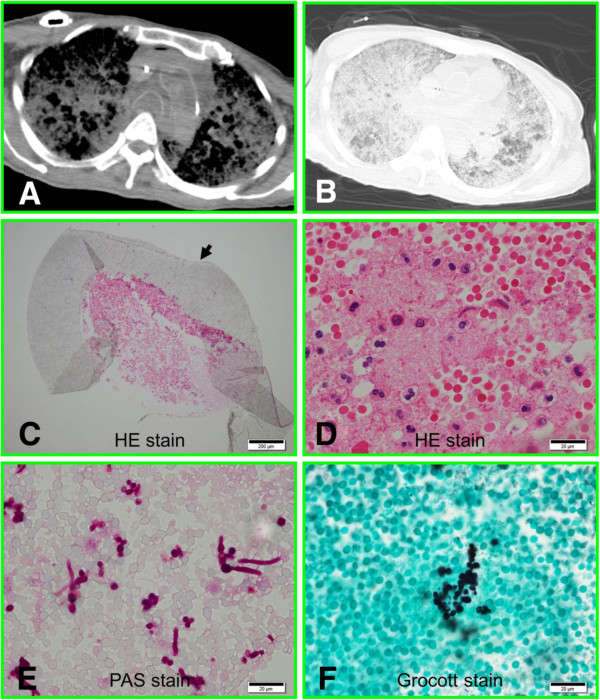
A 62-year-old Japanese female with obstructive jaundice, gastrointestinal bleeding, and liver metastasis from pancreatic head cancer was given an implantable subcutaneous central venous port for nutrition and chemotherapy administration. High fever ensued on day 16 after the central venous port insertion and blood cultures revealed Candida albicans. Although the patient was given 300 mg/day of fosfluconazole according to the suggestion of the infection control team, she died from respiratory failure. Postmortem computed tomography revealed findings consistent with acute respiratory distress syndrome, suggesting that the patient's course was complicated by catheter-related sepsis. Autopsy revealed a subcutaneous abscess around the port, from which C. albicans was cultured. However, no catheter-adherent thrombus, thrombosis of the great central veins, or endocardial vegetations were detected in the patient. Histological analysis revealed scattered abscesses in several organs including lungs and kidneys. Hyaline membrane formation and Candida colonies were found in the lungs. The central venous port tube, together with the part of the subclavian vein into which it had been inserted, was involved in an intraluminal fibrin thrombus containing neutrophils and macrophages, indicating that the thrombus existed while the patient was alive. Histopathological examination following use of the periodic acid-Schiff reagent and the Grocott stain revealed scattered Candida in the thrombus.
Prophylactic thrombolysis should be encouraged to prevent central venous catheter-related candidiasis in clinical practice.
中心静脉导管为静脉输液和营养支持提供了便捷途径,但可能引发诸如导管相关感染等并发症。感染的中心静脉导管常导致医院获得性血流感染,发病率和死亡率较高。然而,已发表的大多数形态学数据来源于体外和体内研究,鲜有关于从患者样本中获取直接证据的报道。在此,我们展示了导管相关念珠菌血症的可视化证据。据我们所知,这是首例报道的患者中心静脉导管内念珠菌感染腔内血栓的传统组织病理学证据。
一名62岁的日本女性,患有梗阻性黄疸、胃肠道出血以及胰头癌肝转移,因营养支持和化疗需要植入了皮下中心静脉港。在中心静脉港植入后第16天,患者出现高热,血培养显示白色念珠菌感染。尽管根据感染控制团队的建议给予患者300mg/天的氟康唑,但患者最终死于呼吸衰竭。尸检计算机断层扫描显示符合急性呼吸窘迫综合征的表现,提示患者病程因导管相关脓毒症而复杂化。尸检发现中心静脉港周围有皮下脓肿,从中培养出白色念珠菌。然而,患者未检测到导管附着血栓、中心大静脉血栓形成或心内膜赘生物。组织学分析显示包括肺和肾在内的多个器官有散在脓肿。肺部发现透明膜形成和念珠菌菌落。中心静脉港导管及其插入的锁骨下静脉部分,存在含有中性粒细胞和巨噬细胞的腔内纤维蛋白血栓,表明血栓在患者生前就已存在。使用过碘酸希夫试剂和格罗科特染色后的组织病理学检查显示血栓中有散在的念珠菌。
在临床实践中,应鼓励预防性溶栓以预防中心静脉导管相关念珠菌病。