Lin Hui, Qu Yangluowa, Geng Zhixin, Li Cheng, Wu Huping, Dong Nuo, Liu Zuguo, Li Wei
Eye Institute of Xiamen University, Xiamen, Fujian, China ; Fujian Provincial Key Laboratory of Ophthalmology and Visual Science, Xiamen, Fujian, China.
Eye Institute of Xiamen University, Xiamen, Fujian, China ; Xiamen University affiliated Xiamen Eye Center, Xiamen, Fujian, China.
PLoS One. 2014 Jan 31;9(1):e87368. doi: 10.1371/journal.pone.0087368. eCollection 2014.
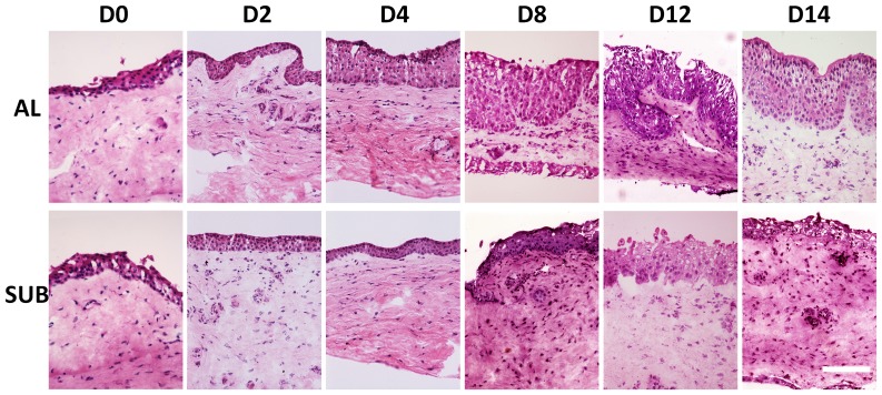
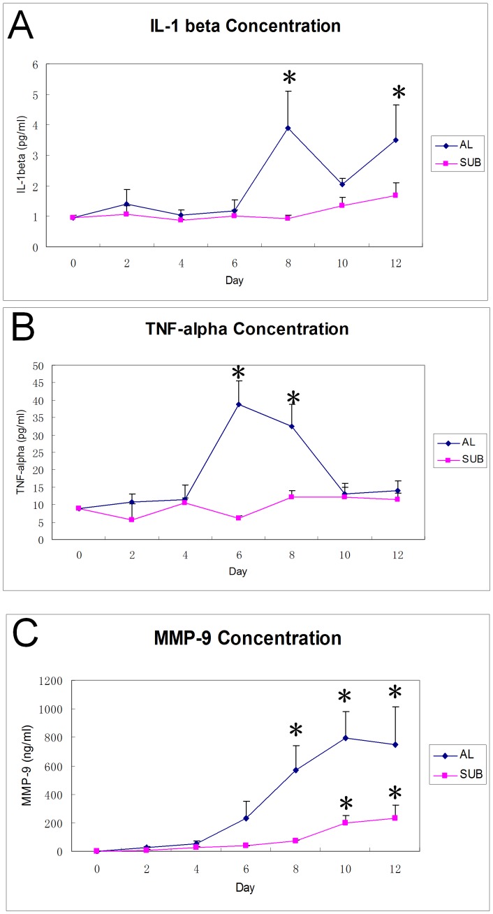
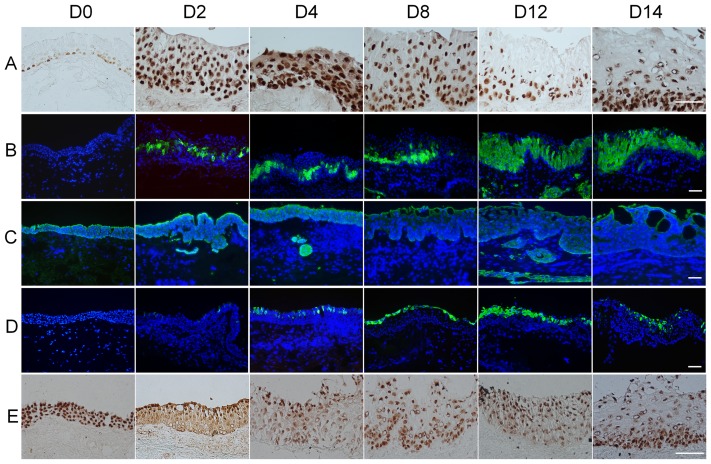
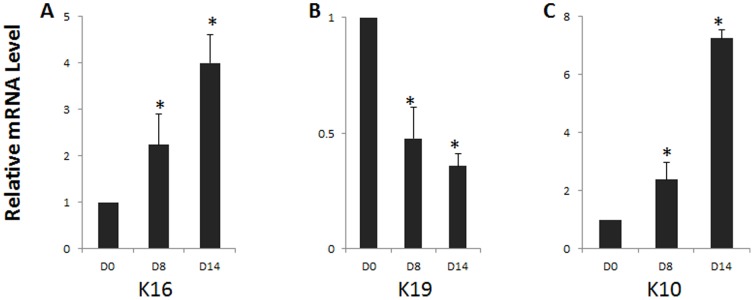
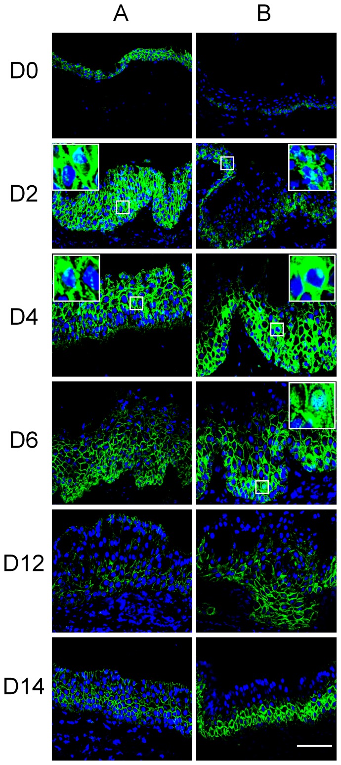
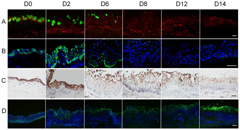
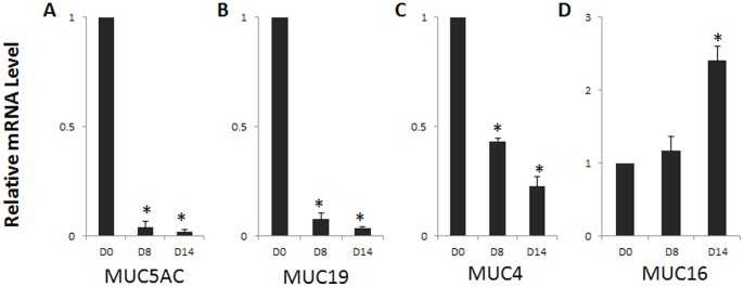
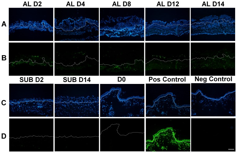
There are several animal models illustrating dry eye pathophysiology. Current study would like to establish an ex vivo tissue culture model for characterizing dry eye. Human conjunctival explants were cultured under airlift or submerged conditions for up to 2 weeks, and only airlifted conjunctival cultures underwent increased epithelial stratification. Starting on day 4, the suprabasal cells displayed decreased K19 expression whereas K10 keratin became evident in airlift group. Pax6 nuclear expression attenuated already at 2 days, while its perinuclear and cytoplasmic expression gradually increased. MUC5AC and MUC19 expression dramatically decreased whereas the full thickness MUC4 and MUC16 expression pattern disappeared soon after initiating the airlift condition. Real time PCR showed K16, K10 and MUC16 gene up-regulated while K19, MUC5AC, MUC19 and MUC4 down-regulated on day 8 and day 14. On day 2 was the appearance of apoptotic epithelial and stromal cells appeared. The Wnt signaling pathway was transiently activated from day 2 to day 10. The inflammatory mediators IL-1β, TNF-α, and MMP-9 were detected in the conditioned media after 6 to 8 days. In conclusion, airlifted conjunctival tissue cultures demonstrated Wnt signaling pathway activation, coupled with squamous metaplasia, mucin pattern alteration, apoptosis and upregulation of proinflammatory cytokine expression. These changes mimic the pathohistological alterations described in dry eye. This correspondence suggests that insight into the pathophysiology of dry eye may be aided through the use of airlifted conjunctival tissue cultures.
有几种动物模型可用于阐释干眼的病理生理学。当前研究希望建立一种用于表征干眼的离体组织培养模型。人结膜外植体在气升或浸没条件下培养长达2周,只有气升培养的结膜出现上皮分层增加。从第4天开始,基底层以上的细胞K19表达降低,而在气升组中K10角蛋白变得明显。Pax6核表达在第2天就开始减弱,而其核周和胞质表达逐渐增加。MUC5AC和MUC19表达显著降低,而在开始气升条件后不久,全层MUC4和MUC16表达模式消失。实时PCR显示在第8天和第14天,K16、K10和MUC16基因上调,而K19、MUC5AC、MUC19和MUC4下调。在第2天出现凋亡的上皮细胞和基质细胞。从第2天到第10天,Wnt信号通路被短暂激活。6至8天后,在条件培养基中检测到炎症介质IL-1β、TNF-α和MMP-9。总之,气升培养的结膜组织显示Wnt信号通路激活,伴有鳞状化生、粘蛋白模式改变、细胞凋亡和促炎细胞因子表达上调。这些变化模拟了干眼中描述的病理组织学改变。这种对应关系表明,通过使用气升培养的结膜组织,可能有助于深入了解干眼的病理生理学。