Department of Geriatrics, Tainan Hospital, Ministry of Health and Welfare, Tainan, Taiwan ; Graduate Institute of Biomedical Electronics and Bioinformatics, National Taiwan University, Taipei, Taiwan.
Research Center for Adaptive Data Analysis, National Central University, Jongli, Taiwan.
PLoS One. 2014 Feb 3;9(2):e87798. doi: 10.1371/journal.pone.0087798. eCollection 2014.
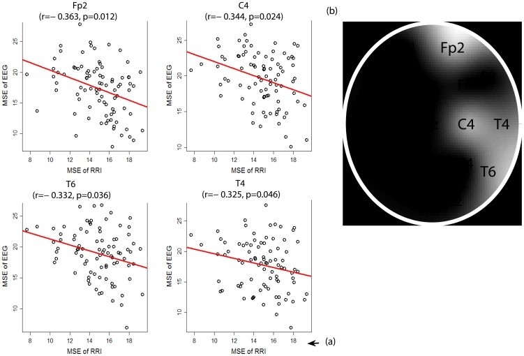
The heart begins to beat before the brain is formed. Whether conventional hierarchical central commands sent by the brain to the heart alone explain all the interplay between these two organs should be reconsidered. Here, we demonstrate correlations between the signal complexity of brain and cardiac activity. Eighty-seven geriatric outpatients with healthy hearts and varied cognitive abilities each provided a 24-hour electrocardiography (ECG) and a 19-channel eye-closed routine electroencephalography (EEG). Multiscale entropy (MSE) analysis was applied to three epochs (resting-awake state, photic stimulation of fast frequencies (fast-PS), and photic stimulation of slow frequencies (slow-PS)) of EEG in the 1-58 Hz frequency range, and three RR interval (RRI) time series (awake-state, sleep and that concomitant with the EEG) for each subject. The low-to-high frequency power (LF/HF) ratio of RRI was calculated to represent sympatho-vagal balance. With statistics after Bonferroni corrections, we found that: (a) the summed MSE value on coarse scales of the awake RRI (scales 11-20, RRI-MSE-coarse) were inversely correlated with the summed MSE value on coarse scales of the resting-awake EEG (scales 6-20, EEG-MSE-coarse) at Fp2, C4, T6 and T4; (b) the awake RRI-MSE-coarse was inversely correlated with the fast-PS EEG-MSE-coarse at O1, O2 and C4; (c) the sleep RRI-MSE-coarse was inversely correlated with the slow-PS EEG-MSE-coarse at Fp2; (d) the RRI-MSE-coarse and LF/HF ratio of the awake RRI were correlated positively to each other; (e) the EEG-MSE-coarse at F8 was proportional to the cognitive test score; (f) the results conform to the cholinergic hypothesis which states that cognitive impairment causes reduction in vagal cardiac modulation; (g) fast-PS significantly lowered the EEG-MSE-coarse globally. Whether these heart-brain correlations could be fully explained by the central autonomic network is unknown and needs further exploration.
心脏在大脑形成之前就开始跳动。大脑向心脏发出的传统分层中枢指令是否能够完全解释这两个器官之间的相互作用,这一点值得重新考虑。在这里,我们展示了大脑和心脏活动信号复杂性之间的相关性。87 名患有健康心脏和不同认知能力的老年门诊患者每人提供了 24 小时心电图(ECG)和 19 通道闭眼常规脑电图(EEG)。多尺度熵(MSE)分析应用于 EEG 在 1-58 Hz 频率范围内的三个时段(休息-清醒状态、快频闪光刺激(fast-PS)和慢频闪光刺激(slow-PS)),以及每个受试者的三个 RR 间隔(RRI)时间序列(清醒状态、睡眠和与 EEG 同时发生的状态)。RR 间隔的低到高频功率(LF/HF)比用于代表交感神经-迷走神经平衡。经过 Bonferroni 校正后的统计学分析,我们发现:(a)清醒 RRI 粗尺度上的总和 MSE 值(尺度 11-20,RRI-MSE-coarse)与休息-清醒 EEG 粗尺度上的总和 MSE 值(尺度 6-20,EEG-MSE-coarse)呈负相关,在 Fp2、C4、T6 和 T4 处;(b)清醒 RRI-MSE-coarse 与快 PS EEG-MSE-coarse 在 O1、O2 和 C4 处呈负相关;(c)睡眠 RRI-MSE-coarse 与慢 PS EEG-MSE-coarse 在 Fp2 处呈负相关;(d)清醒 RRI-MSE-coarse 和清醒 RRI 的 LF/HF 比彼此呈正相关;(e)F8 处的 EEG-MSE-coarse 与认知测试分数成正比;(f)结果符合胆碱能假说,即认知障碍导致迷走神经心脏调节减少;(g)快 PS 显著降低了整体 EEG-MSE-coarse。这些心脏-大脑相关性是否可以完全用自主中枢网络来解释尚不清楚,需要进一步探索。