Granlund Kristin L, Staroswiecki Ernesto, Alley Marcus T, Daniel Bruce L, Hargreaves Brian A
Radiology, Stanford University, Stanford, CA 94305; Electrical Engineering, Stanford University, Stanford, CA 94305.
Radiology, Stanford University, Stanford, CA 94305; Electrical Engineering, Stanford University, Stanford, CA 94305.
Magn Reson Imaging. 2014 May;32(4):330-41. doi: 10.1016/j.mri.2013.12.014. Epub 2014 Jan 3.
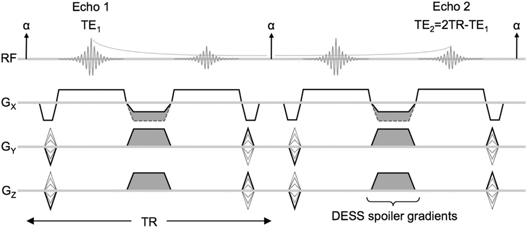
To evaluate the use of the double-echo steady-state (DESS) sequence for acquiring high-resolution breast images with diffusion and T2 weighting.
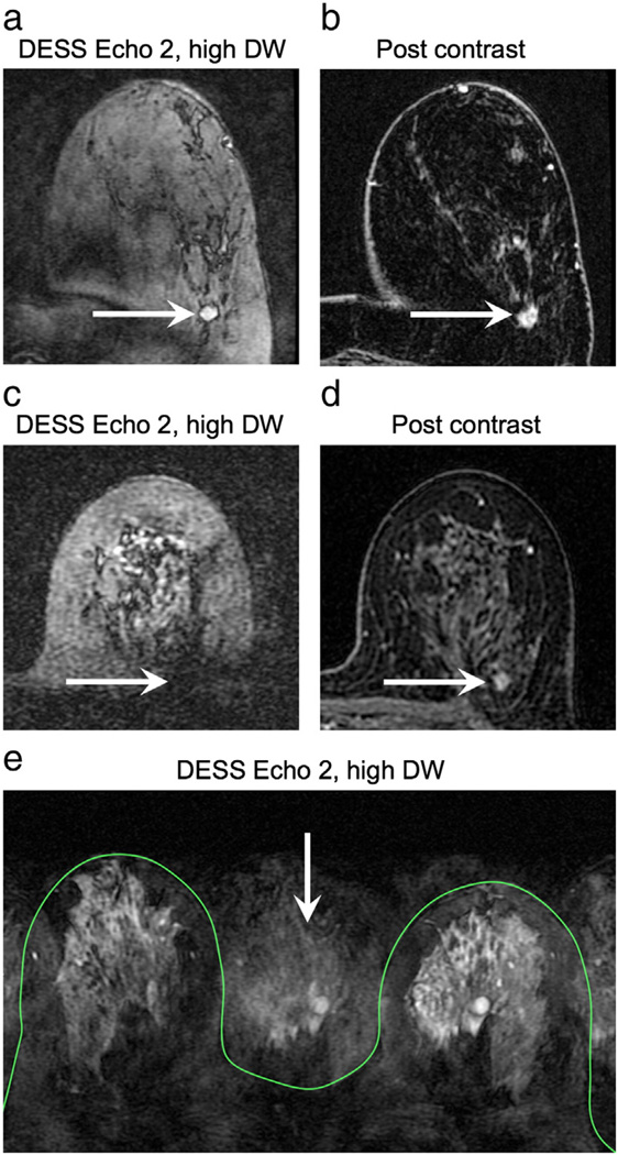
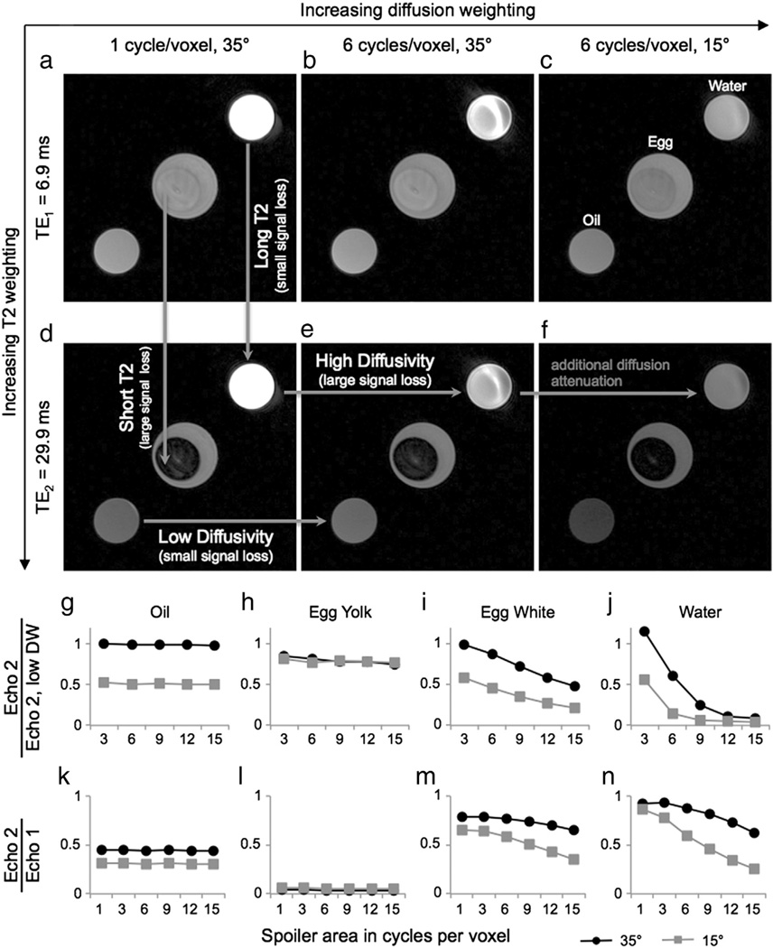
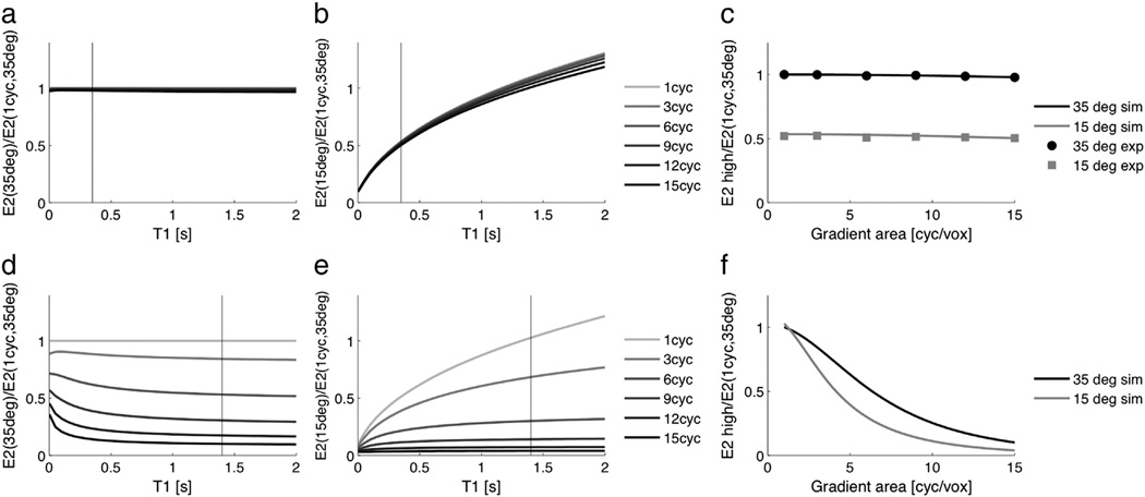
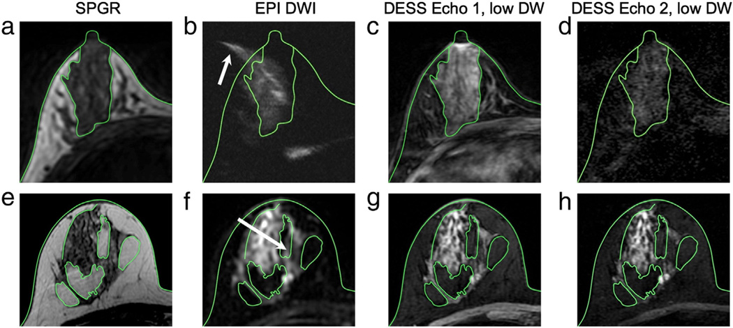
Phantom scans were used to verify the T2 and diffusion weighting of the DESS sequence. Image distortion was evaluated in volunteers by comparing DESS images and conventional diffusion-weighted images (DWI) to spoiled gradient-echo images. The DESS sequence was added to a standard clinical protocol, and the resulting patient images were used to evaluate overall image quality and image contrast in lesions.
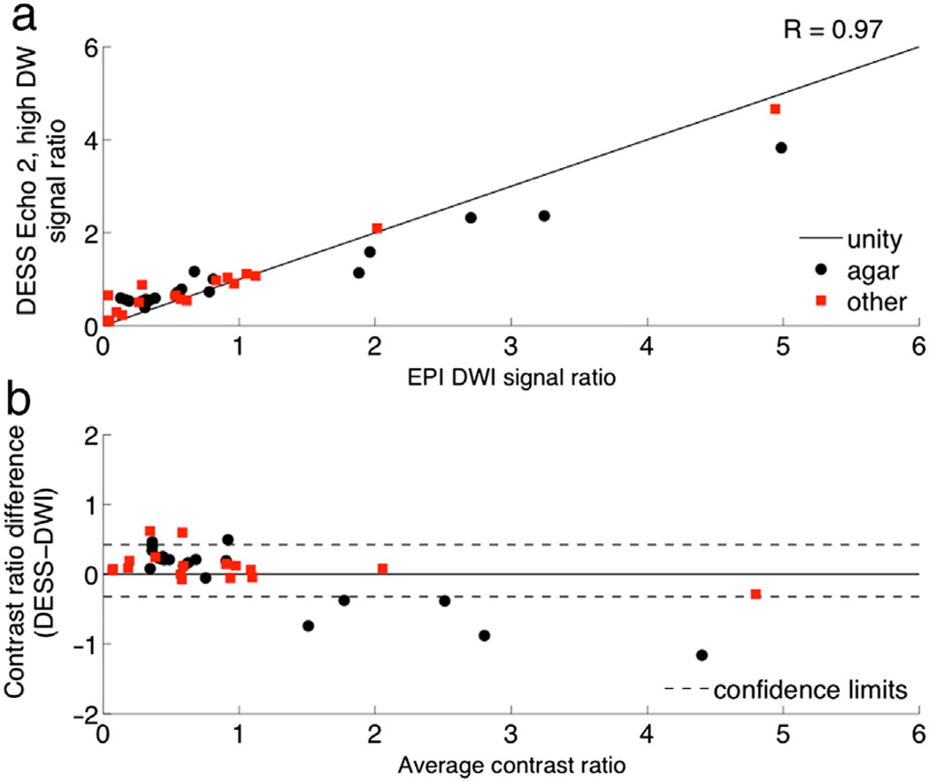
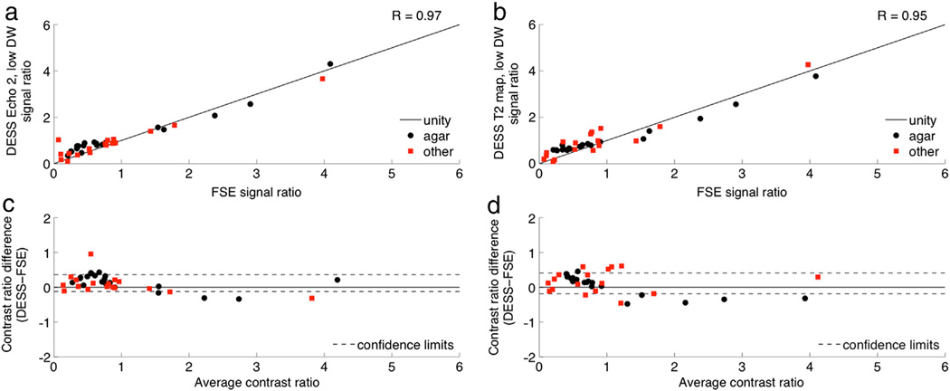
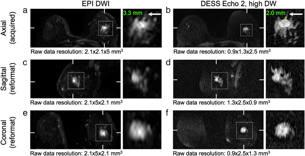
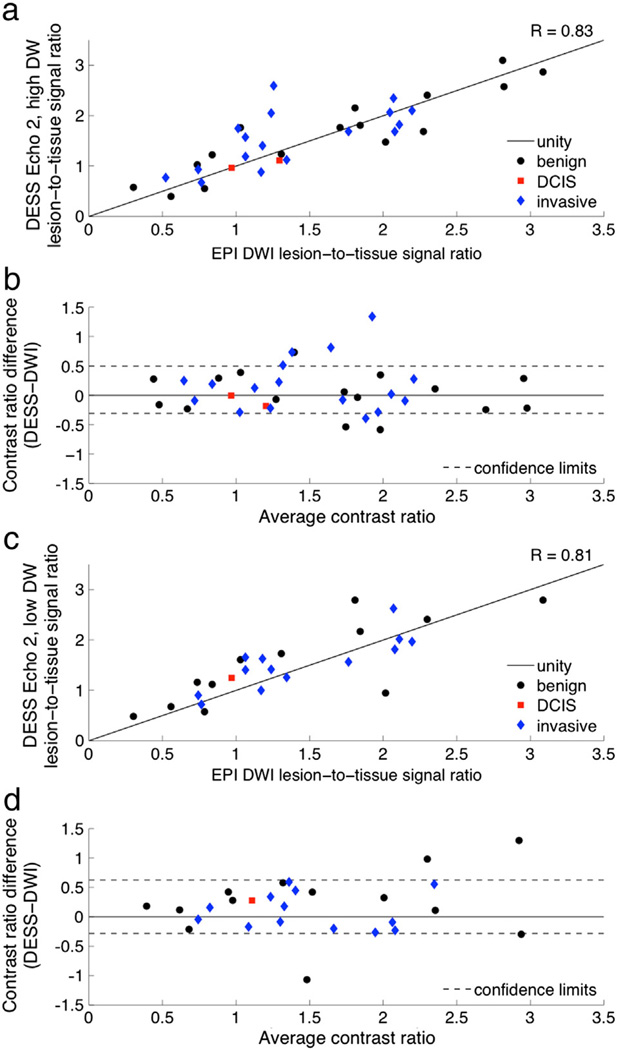
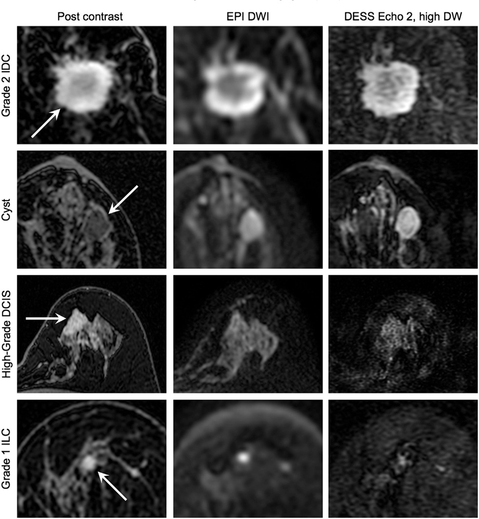
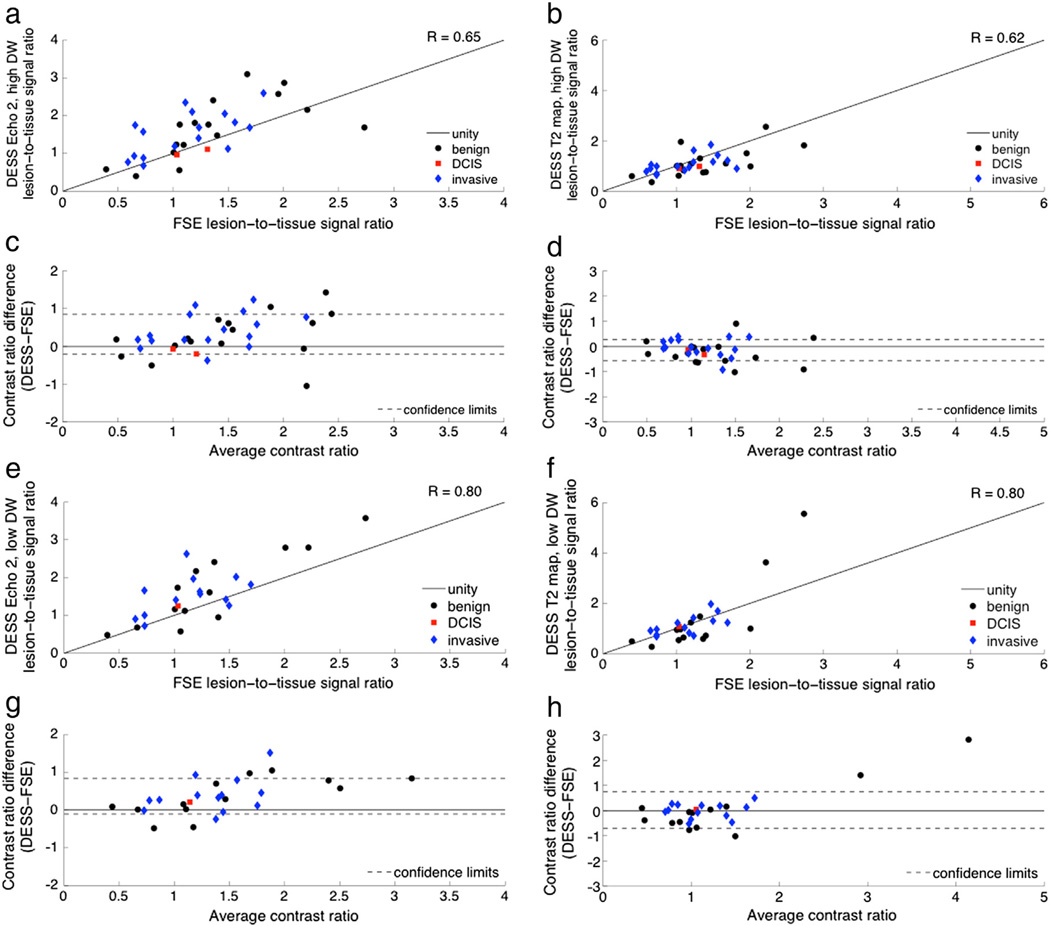
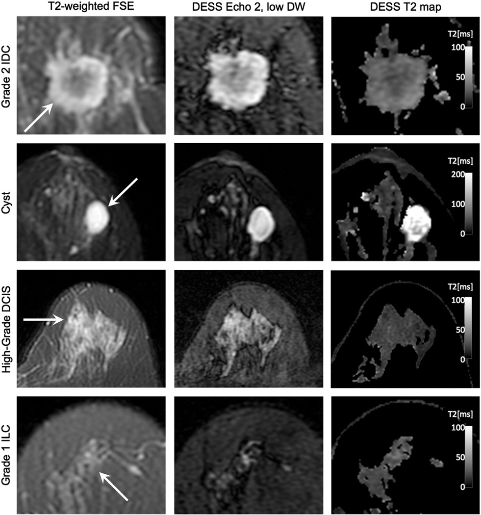
The diffusion weighting of the DESS sequence can be easily modulated by changing the spoiler gradient area and flip angle. Radiologists rated DESS images as having higher resolution and less distortion than conventional DWI. Lesion-to-tissue contrast ratios are strongly correlated between DWI and DESS images (R=0.83) and between T2-weighted fast spin-echo and DESS images (R=0.80).
The DESS sequence is able to acquire high-resolution 3D diffusion- and T2-weighted images in short scan times, with image quality that facilitates morphological assessment of lesions.
评估双回波稳态(DESS)序列在获取具有扩散加权和T2加权的高分辨率乳腺图像中的应用。
使用体模扫描来验证DESS序列的T2和扩散加权。通过将DESS图像和传统扩散加权图像(DWI)与扰相梯度回波图像进行比较,在志愿者中评估图像畸变。将DESS序列添加到标准临床方案中,并使用所得患者图像来评估病变的整体图像质量和图像对比度。
通过改变扰相梯度面积和翻转角,可以轻松调节DESS序列的扩散加权。放射科医生将DESS图像评为比传统DWI具有更高的分辨率和更少的畸变。DWI与DESS图像之间(R = 0.83)以及T2加权快速自旋回波与DESS图像之间(R = 0.80)的病变与组织对比度比值高度相关。
DESS序列能够在短扫描时间内获取高分辨率的三维扩散加权和T2加权图像,其图像质量有助于对病变进行形态学评估。