Richardson Owen C, Bane Octavia, Scott Marietta L J, Tanner Steven F, Waterton John C, Sourbron Steven P, Carroll Timothy J, Buckley David L
Division of Medical Physics, University of Leeds, Leeds, United Kingdom.
Departments of Biomedical Engineering and Radiology, Northwestern University, Chicago, Illinois, USA.
Magn Reson Med. 2015 Jan;73(1):244-53. doi: 10.1002/mrm.25128. Epub 2014 Feb 11.
There is currently no adequate method of mapping physiologic and pathophysiologic tissue albumin concentrations in human subjects. The objective of this study was to devise and evaluate a biomarker of regional albumin concentration using gadofosveset-enhanced MRI.
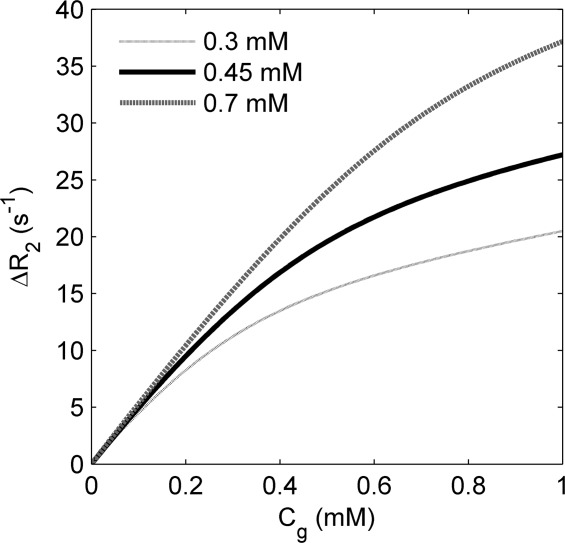
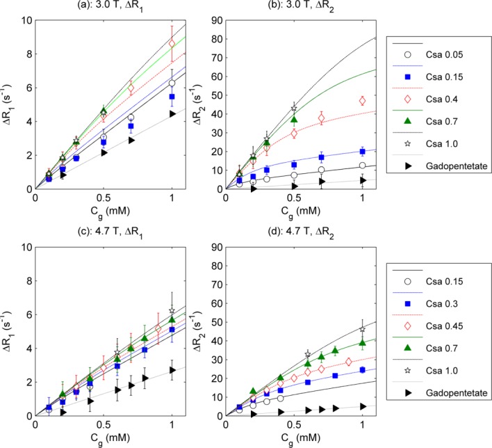
A binding and relaxation model was devised and evaluated in vitro in solutions of albumin at 3.0 Tesla (T) and 4.7T. The method was evaluated in the heart in seven volunteers at 3.0T.
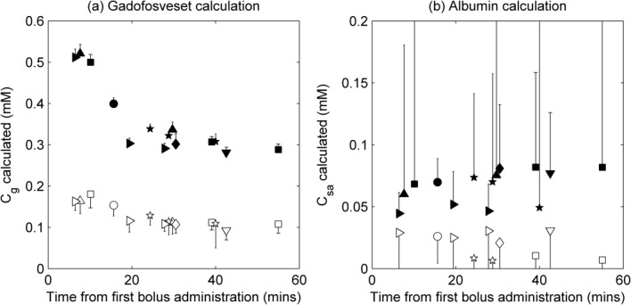
MRI-derived estimates of albumin concentration were in good agreement with true values over the range 0.1-1.0 mM (Pearson correlation coefficients of 0.85 and 0.88 for 3.0T and 4.7T, respectively). The mean calculated albumin concentration in the myocardium for the volunteers was 0.02 mM (range, 0.01-0.03 mM).
Accurate estimates of albumin concentration in vitro suggest this may be a viable noninvasive alternative to existing techniques. In the myocardium the MRI-derived estimates of albumin concentration indicate the practical feasibility of the technique but were below expected values. Gadofosveset-enhanced MR relaxometry has potential in providing biomarkers of regional albumin concentration; further evaluation is required before it can be used reliably in vivo.
目前尚无足够的方法来绘制人体生理和病理生理组织白蛋白浓度图。本研究的目的是利用钆布醇增强磁共振成像(MRI)设计并评估一种区域白蛋白浓度生物标志物。
设计了一种结合与弛豫模型,并在3.0特斯拉(T)和4.7T的白蛋白溶液中进行体外评估。该方法在7名志愿者的心脏中于3.0T下进行了评估。
MRI得出的白蛋白浓度估计值与0.1 - 1.0 mM范围内的真实值高度一致(3.0T和4.7T时的Pearson相关系数分别为0.85和0.88)。志愿者心肌中计算出的白蛋白浓度平均值为0.02 mM(范围为0.01 - 0.03 mM)。
体外白蛋白浓度的准确估计表明这可能是现有技术可行的非侵入性替代方法。在心肌中,MRI得出的白蛋白浓度估计值表明了该技术的实际可行性,但低于预期值。钆布醇增强磁共振弛豫测量法在提供区域白蛋白浓度生物标志物方面具有潜力;在能够可靠地用于体内之前,还需要进一步评估。