• 文献检索
  • 文档翻译
  • 深度研究
  • 学术资讯
  • Suppr Zotero 插件Zotero 插件
  • 邀请有礼
  • 套餐&价格
  • 历史记录
应用&插件
Suppr Zotero 插件Zotero 插件浏览器插件Mac 客户端Windows 客户端微信小程序
定价
高级版会员购买积分包购买API积分包
服务
文献检索文档翻译深度研究API 文档MCP 服务
关于我们
关于 Suppr公司介绍联系我们用户协议隐私条款
关注我们

Suppr 超能文献

核心技术专利:CN118964589B侵权必究
粤ICP备2023148730 号-1Suppr @ 2026

文献检索

告别复杂PubMed语法,用中文像聊天一样搜索,搜遍4000万医学文献。AI智能推荐,让科研检索更轻松。

立即免费搜索

文件翻译

保留排版,准确专业,支持PDF/Word/PPT等文件格式,支持 12+语言互译。

免费翻译文档

深度研究

AI帮你快速写综述,25分钟生成高质量综述,智能提取关键信息,辅助科研写作。

立即免费体验

利用高性能低磁场强度 MRI 进行介入性和诊断性成像的机会。

Opportunities in Interventional and Diagnostic Imaging by Using High-Performance Low-Field-Strength MRI.

机构信息

From the Cardiovascular Branch, Division of Intramural Research, National Heart, Lung, and Blood Institute, National Institutes of Health, Bethesda, Md (A.E.C.W., R.R., M.C.R., I.B., B.B., D.A.H., M.S.H., T.R., W.P.B., D.R.M., C.M., M.Y.C., R.J.L.); Siemens Healthcare GmbH, Erlangen, Germany (D.G., R.S.); Siemens Medical Solutions Inc, Malvern Pa (W.M., H.B.); Systems Biology Center, Division of Intramural Research, National Heart, Lung, and Blood Institute, National Institutes of Health, 10 Center Dr, Building 10, Room 4C-1581, Bethesda, MD 20892-1458 (H.X., P.K., R.S.B.); Pulmonary Branch, Division of Intramural Research, National Heart, Lung, and Blood Institute, National Institutes of Health, Bethesda, MD (J.M.); Department of Radiology and Imaging Sciences, Clinical Center, National Institutes of Health, Bethesda, Md (A.A.M., E.C.J.); and Laboratory of Functional and Molecular Imaging, Division of Intramural Research, National Institute of Neurologic Disorders and Stroke, National Institutes of Health, Bethesda, Md (A.P.K.).

出版信息

Radiology. 2019 Nov;293(2):384-393. doi: 10.1148/radiol.2019190452. Epub 2019 Oct 1.

DOI:10.1148/radiol.2019190452
PMID:31573398
原文链接:https://pmc.ncbi.nlm.nih.gov/articles/PMC6823617/
Abstract

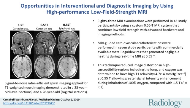
Background Commercial low-field-strength MRI systems are generally not equipped with state-of-the-art MRI hardware, and are not suitable for demanding imaging techniques. An MRI system was developed that combines low field strength (0.55 T) with high-performance imaging technology. Purpose To evaluate applications of a high-performance low-field-strength MRI system, specifically MRI-guided cardiovascular catheterizations with metallic devices, diagnostic imaging in high-susceptibility regions, and efficient image acquisition strategies. Materials and Methods A commercial 1.5-T MRI system was modified to operate at 0.55 T while maintaining high-performance hardware, shielded gradients (45 mT/m; 200 T/m/sec), and advanced imaging methods. MRI was performed between January 2018 and April 2019. T1, T2, and T2* were measured at 0.55 T; relaxivity of exogenous contrast agents was measured; and clinical applications advantageous at low field were evaluated. Results There were 83 0.55-T MRI examinations performed in study participants (45 women; mean age, 34 years ± 13). On average, T1 was 32% shorter, T2 was 26% longer, and T2* was 40% longer at 0.55 T compared with 1.5 T. Nine metallic interventional devices were found to be intrinsically safe at 0.55 T (<1°C heating) and MRI-guided right heart catheterization was performed in seven study participants with commercial metallic guidewires. Compared with 1.5 T, reduced image distortion was shown in lungs, upper airway, cranial sinuses, and intestines because of improved field homogeneity. Oxygen inhalation generated lung signal enhancement of 19% ± 11 (standard deviation) at 0.55 T compared with 7.6% ± 6.3 at 1.5 T ( = .02; five participants) because of the increased T1 relaxivity of oxygen (4.7e-4 mmHgsec). Efficient spiral image acquisitions were amenable to low field strength and generated increased signal-to-noise ratio compared with Cartesian acquisitions ( < .02). Representative imaging of the brain, spine, abdomen, and heart generated good image quality with this system. Conclusion This initial study suggests that high-performance low-field-strength MRI offers advantages for MRI-guided catheterizations with metal devices, MRI in high-susceptibility regions, and efficient imaging. © RSNA, 2019 See also the editorial by Grist in this issue.

摘要

背景 商业低场强 MRI 系统通常不配备最先进的 MRI 硬件,也不适合要求苛刻的成像技术。我们开发了一种将低场强(0.55 T)与高性能成像技术相结合的 MRI 系统。目的 评估高性能低场强 MRI 系统的应用,特别是带有金属器械的 MRI 引导心血管导管术、高磁敏感性区域的诊断成像以及高效的图像采集策略。材料与方法 将商业 1.5 T MRI 系统改装为 0.55 T 运行,同时保持高性能硬件、屏蔽梯度(45 mT/m;200 T/m/sec)和先进的成像方法。MRI 于 2018 年 1 月至 2019 年 4 月进行。在 0.55 T 下测量 T1、T2 和 T2*;测量外源性对比剂的弛豫率;评估在低场具有优势的临床应用。结果 在研究参与者中进行了 83 次 0.55-T MRI 检查(45 名女性;平均年龄 34 岁±13 岁)。与 1.5 T 相比,0.55 T 下 T1 缩短 32%,T2 延长 26%,T2*延长 40%。9 种金属介入器械在 0.55 T 下被证明是安全的(<1°C 加热),并对 7 名研究参与者使用商业金属导丝进行了 MRI 引导的右心导管术。与 1.5 T 相比,由于场均匀性的改善,肺部、上呼吸道、颅窦和肠道的图像失真减少。0.55 T 下吸入氧气使肺部信号增强 19%±11(标准差),而 1.5 T 下为 7.6%±6.3(=.02;5 名参与者),因为氧气的 T1 弛豫率(4.7e-4 mmHgsec)增加。与笛卡尔采集相比,高效螺旋图像采集适用于低场强,可产生更高的信噪比(<.02)。该系统生成的大脑、脊柱、腹部和心脏的代表性成像具有良好的图像质量。结论 这项初步研究表明,高性能低场强 MRI 为 MRI 引导的金属器械导管术、高磁敏感性区域的 MRI 和高效成像提供了优势。

https://cdn.ncbi.nlm.nih.gov/pmc/blobs/4da2/6823617/9e79f52af09c/radiol.2019190452.VA.jpg

相似文献

1
Opportunities in Interventional and Diagnostic Imaging by Using High-Performance Low-Field-Strength MRI.利用高性能低磁场强度 MRI 进行介入性和诊断性成像的机会。
Radiology. 2019 Nov;293(2):384-393. doi: 10.1148/radiol.2019190452. Epub 2019 Oct 1.
2
Susceptibility artifacts from metallic markers and cardiac catheterization devices on a high-performance 0.55 T MRI system.高性能 0.55T MRI 系统上的金属标记物和心导管设备的磁化率伪影。
Magn Reson Imaging. 2021 Apr;77:14-20. doi: 10.1016/j.mri.2020.12.002. Epub 2020 Dec 9.
3
Right heart catheterization using metallic guidewires and low SAR cardiovascular magnetic resonance fluoroscopy at 1.5 Tesla: first in human experience.1.5T 下使用金属导丝和低 SAR 心血管磁共振透视进行右心导管检查:首例人体经验。
J Cardiovasc Magn Reson. 2018 Jun 21;20(1):41. doi: 10.1186/s12968-018-0458-7.
4
Segmented nitinol guidewires with stiffness-matched connectors for cardiovascular magnetic resonance catheterization: preserved mechanical performance and freedom from heating.用于心血管磁共振导管插入术的具有刚度匹配连接器的分段镍钛诺导丝:保留机械性能且无发热问题。
J Cardiovasc Magn Reson. 2015 Nov 30;17:105. doi: 10.1186/s12968-015-0210-5.
5
Positive contrast spiral imaging for visualization of commercial nitinol guidewires with reduced heating.用于可视化商用镍钛诺导丝并减少发热的阳性对比剂螺旋成像
J Cardiovasc Magn Reson. 2015 Dec 22;17:114. doi: 10.1186/s12968-015-0219-9.
6
Whole-Brain Functional and Diffusion Tensor MRI in Human Participants with Metallic Orthodontic Braces.金属正畸牙套对人体全脑功能和弥散张量 MRI 的影响
Radiology. 2020 Jan;294(1):149-157. doi: 10.1148/radiol.2019190070. Epub 2019 Nov 12.
7
Multisequence 3-T Image Synthesis from 64-mT Low-Field-Strength MRI Using Generative Adversarial Networks in Multiple Sclerosis.基于生成对抗网络的多序列3-T图像从64-mT低场强磁共振成像合成在多发性硬化症中的应用
Radiology. 2025 Apr;315(1):e233529. doi: 10.1148/radiol.233529.
8
Modern Low-Field MRI of the Musculoskeletal System: Practice Considerations, Opportunities, and Challenges.现代肌肉骨骼系统低场磁共振成像:实践考量、机遇与挑战
Invest Radiol. 2023 Jan 1;58(1):76-87. doi: 10.1097/RLI.0000000000000912. Epub 2022 Sep 13.
9
MRI for Guided Right and Left Heart Cardiac Catheterization: A Prospective Study in Congenital Heart Disease.MRI 引导下的右心和左心心导管检查:先天性心脏病的前瞻性研究。
J Magn Reson Imaging. 2021 May;53(5):1446-1457. doi: 10.1002/jmri.27426. Epub 2020 Nov 6.
10
Instrument visualization using conventional and compressed sensing SEMAC for interventional MRI at 3T.使用常规和压缩感知 SEMAC 对 3T 介入 MRI 进行器械可视化。
J Magn Reson Imaging. 2018 May;47(5):1306-1315. doi: 10.1002/jmri.25858. Epub 2017 Sep 21.

引用本文的文献

1
4D-MRI at 0.55T for internal target volume delineation in liver radiation therapy planning.用于肝脏放射治疗计划中内部靶区勾画的0.55T 4D-MRI
Med Phys. 2025 Aug;52(8):e18069. doi: 10.1002/mp.18069.
2
SNRAware: Improved Deep Learning MRI Denoising with SNR Unit Training and G-factor Map Augmentation.SNRAware:通过信噪比单元训练和G因子图增强改进深度学习磁共振成像去噪
ArXiv. 2025 Mar 23:arXiv:2503.18162v1.
3
High-resolution 3D whole-heart bright- and black-blood imaging with co-registered T2 mapping at 0.55 T.在0.55 T磁场下进行高分辨率3D全心亮血与黑血成像及T2映射配准

本文引用的文献

1
Low-field MRI: An MR physics perspective.低场 MRI:磁共振物理视角。
J Magn Reson Imaging. 2019 Jun;49(6):1528-1542. doi: 10.1002/jmri.26637. Epub 2019 Jan 13.
2
Cardiac balanced steady-state free precession MRI at 0.35 T: a comparison study with 1.5 T.0.35T心脏平衡稳态自由进动磁共振成像:与1.5T的比较研究
Quant Imaging Med Surg. 2018 Aug;8(7):627-636. doi: 10.21037/qims.2018.08.09.
3
Dynamic off-resonance correction for spiral real-time MRI of speech.语音螺旋实时 MRI 的动态离频校正。
Front Cardiovasc Med. 2025 Jul 3;12:1572318. doi: 10.3389/fcvm.2025.1572318. eCollection 2025.
4
In-vivo liver proton density fat fraction quantification at 0.55 T: a pilot study with comparison against 3 T MRI.0.55T下肝脏质子密度脂肪分数的活体定量:一项与3T MRI对比的初步研究。
MAGMA. 2025 Jul 15. doi: 10.1007/s10334-025-01277-9.
5
Real-Time MR-Guided Lumbosacral Periradicular Injection Therapy Using a 0.55 T MRI System: A Phantom Study.使用0.55 T磁共振成像系统进行实时磁共振引导下腰骶部神经根周围注射治疗:一项体模研究
Diagnostics (Basel). 2025 Jun 2;15(11):1413. doi: 10.3390/diagnostics15111413.
6
Initial experience of cardiac Tρ mapping at 0.55 T: Continuous wave versus adiabatic spin-lock preparation pulses.0.55T心脏Tρ成像的初步经验:连续波与绝热自旋锁定准备脉冲对比
Magn Reson Med. 2025 Oct;94(4):1644-1653. doi: 10.1002/mrm.30582. Epub 2025 May 20.
7
Longitudinal relaxation rate ( ) in lung: a systematic review and meta-analysis.肺的纵向弛豫率( ):一项系统评价和荟萃分析。
MAGMA. 2025 May 16. doi: 10.1007/s10334-025-01259-x.
8
Super-Field MRI Synthesis for Infant Brains Enhanced by Dual Channel Latent Diffusion.双通道潜在扩散增强的婴儿大脑超场磁共振成像合成
Med Image Comput Comput Assist Interv. 2024 Oct;15003:444-454. doi: 10.1007/978-3-031-72384-1_42. Epub 2024 Oct 3.
9
Comparative study of radiofrequency heating in deep brain stimulation devices during MRI at 1.5 T and 0.55 T: Challenging the assumption of safety at low field strengths.1.5T和0.55T磁共振成像期间深部脑刺激装置中射频加热的比较研究:质疑低场强安全性的假设
Magn Reson Med. 2025 Aug;94(2):785-796. doi: 10.1002/mrm.30515. Epub 2025 Apr 28.
10
Contactless cardiac gating at 0.55T using high-amplitude pilot tone with interference cancellation (HAPTIC).使用带干扰消除的高振幅导频音(HAPTIC)在0.55T下进行非接触式心脏门控。
Magn Reson Med. 2025 Sep;94(3):1182-1190. doi: 10.1002/mrm.30528. Epub 2025 Apr 14.
Magn Reson Med. 2019 Jan;81(1):234-246. doi: 10.1002/mrm.27373. Epub 2018 Jul 29.
4
Right heart catheterization using metallic guidewires and low SAR cardiovascular magnetic resonance fluoroscopy at 1.5 Tesla: first in human experience.1.5T 下使用金属导丝和低 SAR 心血管磁共振透视进行右心导管检查:首例人体经验。
J Cardiovasc Magn Reson. 2018 Jun 21;20(1):41. doi: 10.1186/s12968-018-0458-7.
5
Improved passive catheter tracking with positive contrast for CMR-guided cardiac catheterization using partial saturation (pSAT).使用部分饱和(pSAT)技术增强 CMR 引导下心导管术的被动导管跟踪的正对比。
J Cardiovasc Magn Reson. 2017 Aug 15;19(1):60. doi: 10.1186/s12968-017-0368-0.
6
Low-Field Cardiac Magnetic Resonance Imaging: A Compelling Case for Cardiac Magnetic Resonance's Future.低场心脏磁共振成像:心脏磁共振未来发展的有力例证
Circ Cardiovasc Imaging. 2017 Jun;10(6). doi: 10.1161/CIRCIMAGING.117.005446.
7
T1 relaxation time constants, influence of oxygen, and the oxygen transfer function of the human lung at 1.5T-A meta-analysis.1.5T下人体肺脏的T1弛豫时间常数、氧的影响及氧传递函数——一项荟萃分析
Eur J Radiol. 2017 Jan;86:252-260. doi: 10.1016/j.ejrad.2016.11.027. Epub 2016 Nov 24.
8
Lymphangioleiomyomatosis.淋巴管平滑肌瘤病。
Clin Chest Med. 2016 Sep;37(3):389-403. doi: 10.1016/j.ccm.2016.04.002.
9
Cardiovascular MRI with ferumoxytol.使用铁羧麦芽糖的心血管磁共振成像。
Clin Radiol. 2016 Aug;71(8):796-806. doi: 10.1016/j.crad.2016.03.020. Epub 2016 May 21.
10
Safety and technique of ferumoxytol administration for MRI.用于磁共振成像的铁羧麦芽糖给药的安全性与技术
Magn Reson Med. 2016 May;75(5):2107-11. doi: 10.1002/mrm.26151. Epub 2016 Feb 18.