• 文献检索
  • 文档翻译
  • 深度研究
  • 学术资讯
  • Suppr Zotero 插件Zotero 插件
  • 邀请有礼
  • 套餐&价格
  • 历史记录
应用&插件
Suppr Zotero 插件Zotero 插件浏览器插件Mac 客户端Windows 客户端微信小程序
定价
高级版会员购买积分包购买API积分包
服务
文献检索文档翻译深度研究API 文档MCP 服务
关于我们
关于 Suppr公司介绍联系我们用户协议隐私条款
关注我们

Suppr 超能文献

核心技术专利:CN118964589B侵权必究
粤ICP备2023148730 号-1Suppr @ 2026

文献检索

告别复杂PubMed语法,用中文像聊天一样搜索,搜遍4000万医学文献。AI智能推荐,让科研检索更轻松。

立即免费搜索

文件翻译

保留排版,准确专业,支持PDF/Word/PPT等文件格式,支持 12+语言互译。

免费翻译文档

深度研究

AI帮你快速写综述,25分钟生成高质量综述,智能提取关键信息,辅助科研写作。

立即免费体验

小鼠牙髓干细胞向成骨细胞和破骨细胞分化的能力。

Differentiation capacity of mouse dental pulp stem cells into osteoblasts and osteoclasts.

机构信息

School of Biosciences and Biotechnology, Faculty of Science and Technology, University Kebangsaan Malaysia, 43600 Bangi, Selangor, Malaysia.

Department of Orthodontics, Faculty of Dentistry, University Kebangsaan Malaysia, Jalan Raja Muda Abdul Aziz, 50300 Kuala Lumpur, Malaysia.

出版信息

Cell J. 2014 Feb 3;16(1):31-42.

PMID:24518973
原文链接:https://pmc.ncbi.nlm.nih.gov/articles/PMC3933437/
Abstract

OBJECTIVE

Our research attempted to show that mouse dental pulp stem cells (DPSCs) with characters such as accessibility, propagation and higher proliferation rate can provide an improved approach for generate bone tissues. With the aim of finding and comparing the differentiation ability of mesenchymal stem cells derived from DPSCs into osteoblast and osteoclast cells; morphological, molecular and biochemical analyses were conducted.

MATERIALS AND METHODS

In this experimental study, osteoblast and osteoclast differentiation was induced by specific differentiation medium. In order to induce osteoblast differentiation, 50 μg mL(-1) ascorbic acid and 10 mM β-glycerophosphate as growth factors were added to the complete medium consisting alpha-modified Eagle's medium (α-MEM), 15% fetal bovine serum (FBS) and penicillin/streptomycin, while in order to induce the osteoclast differentiation, 10 ng/mL receptor activator of nuclear factor kappa-B ligand (RANKL) and 5 ng/mL macrophage-colony stimulating factor (M-CSF) were added to complete medium. Statistical comparison between the osteoblast and osteoclast differentiated groups and control were carried out using t test.

RESULTS

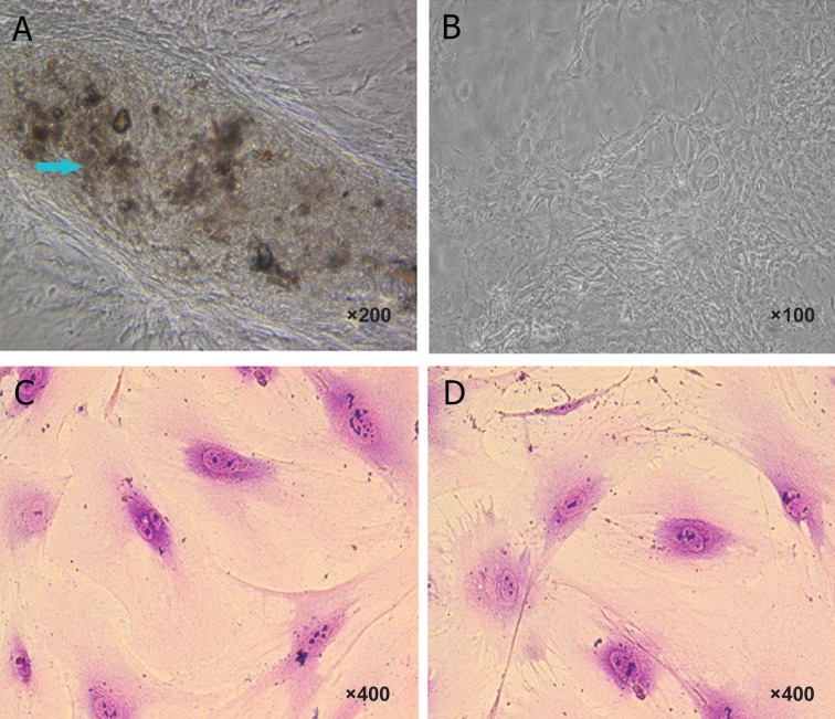
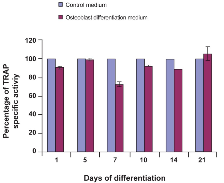
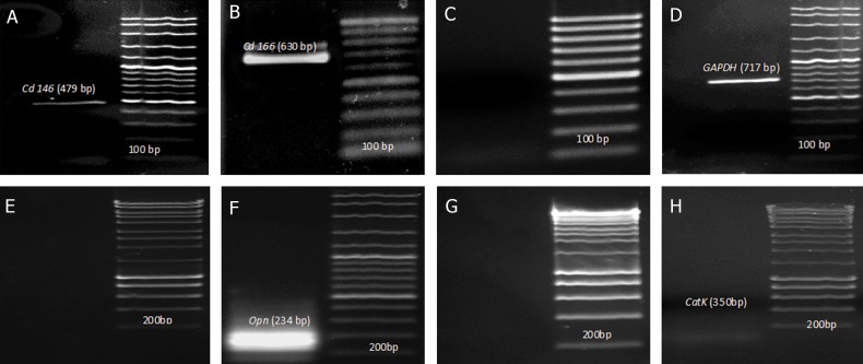
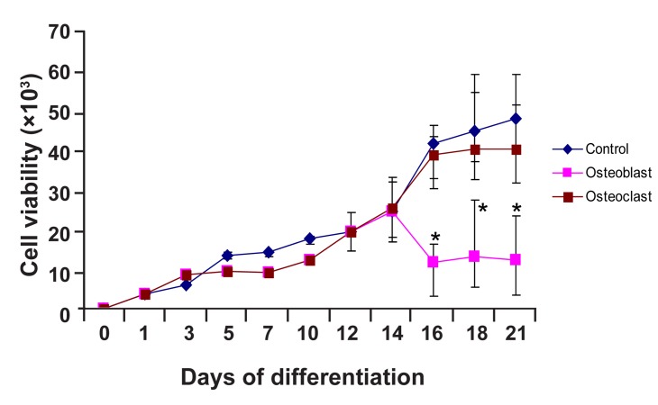
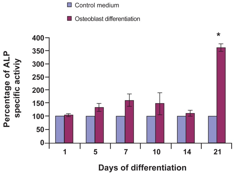
Proliferation activity of cells was estimated by 3-[4,5-dimethylthiazol-2-yl]-2,5 diphenyl tetrazolium bromide (MTT) assay. Statistical results demonstrated significant difference (p<0.05) between the control and osteoblastic induction group, whereas osteoclast cells maintained its proliferation rate (p>0.05). Morphological characterization of osteoblast and osteoclast was evaluated using von Kossa staining and May-Grunwald-Giemsa technique, respectively. Reverse transcription-polymerase chain reaction (RT-PCR) molecular analysis demonstrated that mouse DPSCs expressed Cd146 and Cd166 markers, but did not express Cd31, indicating that these cells belong to mesenchymal stem cells. Osteoblast cells with positive osteopontin (Opn) marker were found after 21 days, whereas this marker was negative for DPSCs. CatK, as an osteoclast marker, was negative in both osteoclast differentiation medium and control group. Biochemical analyses in osteoblast differentiated groups showed alkaline phosphatase (ALP) activity significantly increased on day 21 as compared to control (p<0.05). In osteoclast differentiated groups, tartrate-resistant acid phosphatase (TRAP) activity representing osteoclast biomarker didn't show statistically significant as compared to control (p>0.05).

CONCLUSION

DPSCs have the ability to differentiate into osteoblast, but not into osteoclast.

摘要

目的

我们的研究试图表明,具有易得性、增殖能力和更高增殖率等特征的小鼠牙髓干细胞(DPSCs)可为生成骨组织提供一种改良方法。本研究旨在寻找和比较来源于 DPSCs 的间充质干细胞向成骨细胞和破骨细胞分化的能力;进行了形态学、分子和生化分析。

材料和方法

在这项实验研究中,通过特定的分化培养基诱导成骨细胞和破骨细胞分化。为了诱导成骨细胞分化,在含有α-改良 Eagle 培养基(α-MEM)、15%胎牛血清(FBS)和青霉素/链霉素的完全培养基中加入 50μg/ml 的抗坏血酸和 10mMβ-甘油磷酸作为生长因子,而要诱导破骨细胞分化,则在完全培养基中加入 10ng/ml 核因子κ B 配体受体激活剂(RANKL)和 5ng/ml 巨噬细胞集落刺激因子(M-CSF)。使用 t 检验对成骨细胞和破骨细胞分化组与对照组进行统计学比较。

结果

通过 3-[4,5-二甲基噻唑-2-基]-2,5-二苯基四氮唑溴盐(MTT)测定法估计细胞的增殖活性。统计结果表明,对照组与成骨诱导组之间存在显著差异(p<0.05),而破骨细胞则保持其增殖率(p>0.05)。通过 von Kossa 染色和 May-Grunwald-Giemsa 技术分别评估成骨细胞和破骨细胞的形态特征。逆转录-聚合酶链反应(RT-PCR)分子分析表明,小鼠 DPSCs 表达 Cd146 和 Cd166 标志物,但不表达 Cd31,表明这些细胞属于间充质干细胞。在第 21 天发现具有骨桥蛋白(Opn)标志物阳性的成骨细胞,而 DPSCs 则为阴性。作为破骨细胞标志物的组织蛋白酶 K(CatK)在成骨细胞分化培养基和对照组中均为阴性。在成骨细胞分化组中,碱性磷酸酶(ALP)活性在第 21 天与对照组相比显著增加(p<0.05)。在破骨细胞分化组中,作为破骨细胞生物标志物的抗酒石酸酸性磷酸酶(TRAP)活性与对照组相比没有统计学意义(p>0.05)。

结论

DPSCs 具有分化为成骨细胞的能力,但不能分化为破骨细胞。

https://cdn.ncbi.nlm.nih.gov/pmc/blobs/0dcb/3933437/6c0183e69bc5/Cell-J-16-31-g05.jpg
https://cdn.ncbi.nlm.nih.gov/pmc/blobs/0dcb/3933437/5a4273e557d9/Cell-J-16-31-g01.jpg
https://cdn.ncbi.nlm.nih.gov/pmc/blobs/0dcb/3933437/dae61761115c/Cell-J-16-31-g02.jpg
https://cdn.ncbi.nlm.nih.gov/pmc/blobs/0dcb/3933437/9bfa978c17f8/Cell-J-16-31-g03.jpg
https://cdn.ncbi.nlm.nih.gov/pmc/blobs/0dcb/3933437/7fda9d8afc81/Cell-J-16-31-g04.jpg
https://cdn.ncbi.nlm.nih.gov/pmc/blobs/0dcb/3933437/6c0183e69bc5/Cell-J-16-31-g05.jpg

相似文献

1
Differentiation capacity of mouse dental pulp stem cells into osteoblasts and osteoclasts.小鼠牙髓干细胞向成骨细胞和破骨细胞分化的能力。
Cell J. 2014 Feb 3;16(1):31-42.
2
A Comparative Analysis of Ascorbic Acid-induced Cytotoxicity and Differentiation between SHED and DPSC.抗坏血酸诱导的 SHED 和 DPSC 细胞毒性和分化的比较分析。
Curr Stem Cell Res Ther. 2022;17(6):576-588. doi: 10.2174/1574888X17666220124141310.
3
Aging increases stromal/osteoblastic cell-induced osteoclastogenesis and alters the osteoclast precursor pool in the mouse.衰老会增加基质/成骨细胞诱导的破骨细胞生成,并改变小鼠体内破骨细胞前体细胞库。
J Bone Miner Res. 2005 Sep;20(9):1659-68. doi: 10.1359/JBMR.050503. Epub 2005 May 2.
4
Eluted zinc ions stimulate osteoblast differentiation and mineralization in human dental pulp stem cells for bone tissue engineering.洗脱的锌离子可刺激人牙髓干细胞的成骨细胞分化和矿化,用于骨组织工程。
Arch Oral Biol. 2016 Nov;71:162-169. doi: 10.1016/j.archoralbio.2016.07.010. Epub 2016 Aug 5.
5
Streptococcus gordonii induces bone resorption by increasing osteoclast differentiation and reducing osteoblast differentiation.戈登链球菌通过增加破骨细胞分化和减少成骨细胞分化来诱导骨吸收。
Microb Pathog. 2019 Jan;126:218-223. doi: 10.1016/j.micpath.2018.11.005. Epub 2018 Nov 7.
6
Dental pulp stem cells from traumatically exposed pulps exhibited an enhanced osteogenic potential and weakened odontogenic capacity.外伤性暴露牙髓中的牙髓干细胞表现出增强的成骨潜力和减弱的成牙骨质能力。
Arch Oral Biol. 2013 Nov;58(11):1709-17. doi: 10.1016/j.archoralbio.2013.09.001. Epub 2013 Sep 10.
7
Interleukin-3 plays dual roles in osteoclastogenesis by promoting the development of osteoclast progenitors but inhibiting the osteoclastogenic process.白细胞介素-3 通过促进破骨细胞前体的发育但抑制破骨细胞生成过程在破骨细胞生成中发挥双重作用。
Biochem Biophys Res Commun. 2013 Nov 1;440(4):545-50. doi: 10.1016/j.bbrc.2013.09.098. Epub 2013 Oct 5.
8
Osteogenic properties of human dental pulp stem cells.人牙髓干细胞的成骨特性。
J Biol Regul Homeost Agents. 2010 Apr-Jun;24(2):167-75.
9
Osteoblastic and osteoclastic differentiation on SLA and hydrophilic modified SLA titanium surfaces.在SLA和亲水改性SLA钛表面上的成骨细胞和成破骨细胞分化。
Clin Oral Implants Res. 2014 Jul;25(7):831-7. doi: 10.1111/clr.12146. Epub 2013 Apr 8.
10
[Difference of in vitro osteogenic differentiation and osteoclast capacity between stem cells from human exfoliated deciduous teeth and dental pulp stem cells].人乳牙脱落干细胞与牙髓干细胞在体外成骨分化及破骨细胞能力方面的差异
Nan Fang Yi Ke Da Xue Xue Bao. 2016 Feb;36(2):180-5.

引用本文的文献

1
Dysregulation of lncRNA GATA3-AS1 is Involved in the Pathogenesis of Pulpitis by the Regulation of miR-17-3p.长链非编码RNA GATA3-AS1的失调通过调控miR-17-3p参与牙髓炎的发病机制。
J Inflamm Res. 2025 Mar 26;18:4459-4469. doi: 10.2147/JIR.S504048. eCollection 2025.
2
Comparative evaluation of the osteogenic capacity of second-generation platelet concentrates on dental pulp stem cells - An study.第二代血小板浓缩物对牙髓干细胞成骨能力的比较评估——一项研究。
J Conserv Dent Endod. 2024 Aug;27(8):853-859. doi: 10.4103/JCDE.JCDE_320_24. Epub 2024 Aug 7.
3
Evaluation of osteoblast and osteoclast differentiation from stem cell: a systematic review of morphological assays and staining techniques.

本文引用的文献

1
Determination of the differentiation capacities of murines' primary mononucleated cells and MC3T3-E1 cells.检测鼠源原代单核细胞和 MC3T3-E1 细胞的分化能力。
Cancer Cell Int. 2010 Oct 28;10:42. doi: 10.1186/1475-2867-10-42.
2
Effects of TGF-beta3 and preculture period of osteogenic cells on the chondrogenic differentiation of rabbit marrow mesenchymal stem cells encapsulated in a bilayered hydrogel composite.双层水凝胶复合载体中 TGF-β3 和骨髓间充质干细胞成骨细胞预培养期对其向软骨分化的影响。
Acta Biomater. 2010 Aug;6(8):2920-31. doi: 10.1016/j.actbio.2010.02.046. Epub 2010 Mar 1.
3
Human dental pulp stem cells with highly angiogenic and neurogenic potential for possible use in pulp regeneration.
评价骨髓间充质干细胞向成骨细胞和破骨细胞分化:形态学检测和染色技术的系统评价。
PeerJ. 2024 Jul 25;12:e17790. doi: 10.7717/peerj.17790. eCollection 2024.
4
Comparative evaluation of osteogenic differentiation potential of stem cells derived from dental pulp and exfoliated deciduous teeth cultured over granular hydroxyapatite based scaffold.基于颗粒状羟基磷灰石支架培养的牙髓干细胞与脱落乳牙干细胞成骨分化潜能的比较评估
BMC Oral Health. 2021 May 15;21(1):263. doi: 10.1186/s12903-021-01621-0.
5
Human Dental Pulp Stem Cells Exhibit Osteogenic Differentiation Potential.人牙髓干细胞具有成骨分化潜能。
Open Life Sci. 2020 May 6;15:229-236. doi: 10.1515/biol-2020-0023. eCollection 2020.
6
Is There Such a Thing as a Genuine Cancer Stem Cell Marker? Perspectives from the Gut, the Brain and the Dental Pulp.是否存在真正的癌症干细胞标志物?来自肠道、大脑和牙髓的观点。
Biology (Basel). 2020 Nov 27;9(12):426. doi: 10.3390/biology9120426.
7
Dental pulp stem cells and osteogenesis: an update.牙髓干细胞与骨生成:最新进展
Cytotechnology. 2018 Oct;70(5):1479-1486. doi: 10.1007/s10616-018-0225-5. Epub 2018 Jun 25.
8
Interconnectable Dynamic Compression Bioreactors for Combinatorial Screening of Cell Mechanobiology in Three Dimensions.可互连成组动态压缩生物反应器,用于三维组合筛选细胞机械生物学
ACS Appl Mater Interfaces. 2018 Apr 25;10(16):13293-13303. doi: 10.1021/acsami.7b17991. Epub 2018 Apr 13.
具有高血管生成和神经生成潜力的人牙髓干细胞,可能用于牙髓再生。
Cytokine Growth Factor Rev. 2009 Oct-Dec;20(5-6):435-40. doi: 10.1016/j.cytogfr.2009.10.012. Epub 2009 Nov 6.
4
Effect of ascorbic acid on bone marrow-derived mesenchymal stem cell proliferation and differentiation.抗坏血酸对骨髓间充质干细胞增殖和分化的影响。
J Biosci Bioeng. 2008 Jun;105(6):586-94. doi: 10.1263/jbb.105.586.
5
Osteoblast differentiation of human bone marrow cells under continuous and discontinuous treatment with dexamethasone.地塞米松连续和间断处理下人骨髓细胞的成骨细胞分化
Braz Dent J. 2005;16(2):156-61. doi: 10.1590/s0103-64402005000200013.
6
Comparative characteristics of mesenchymal stem cells from human bone marrow, adipose tissue, and umbilical cord blood.来自人骨髓、脂肪组织和脐带血的间充质干细胞的比较特征。
Exp Hematol. 2005 Nov;33(11):1402-16. doi: 10.1016/j.exphem.2005.07.003.
7
Overexpression of QM induces cell differentiation and mineralization in MC3T3-E1.QM的过表达诱导MC3T3-E1细胞分化和矿化。
Biol Pharm Bull. 2005 Aug;28(8):1371-6. doi: 10.1248/bpb.28.1371.
8
The efficacy of mesenchymal stem cells to regenerate and repair dental structures.间充质干细胞对牙齿结构进行再生和修复的功效。
Orthod Craniofac Res. 2005 Aug;8(3):191-9. doi: 10.1111/j.1601-6343.2005.00331.x.
9
A bio-assay for effectors of osteoclast differentiation in serum from patients with bone disease.
Clin Chim Acta. 2005 Jun;356(1-2):154-63. doi: 10.1016/j.cccn.2005.01.020. Epub 2005 Mar 19.
10
Plasticity of bone marrow-derived stem cells.骨髓源性干细胞的可塑性。
Stem Cells. 2004;22(4):487-500. doi: 10.1634/stemcells.22-4-487.