Fernández de Castro Isabel, Zamora Paula F, Ooms Laura, Fernández José Jesús, Lai Caroline M-H, Mainou Bernardo A, Dermody Terence S, Risco Cristina
mBio. 2014 Feb 18;5(1):e00931-13. doi: 10.1128/mBio.00931-13.
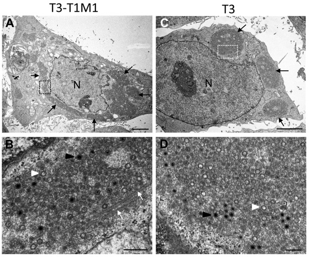
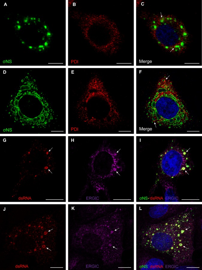
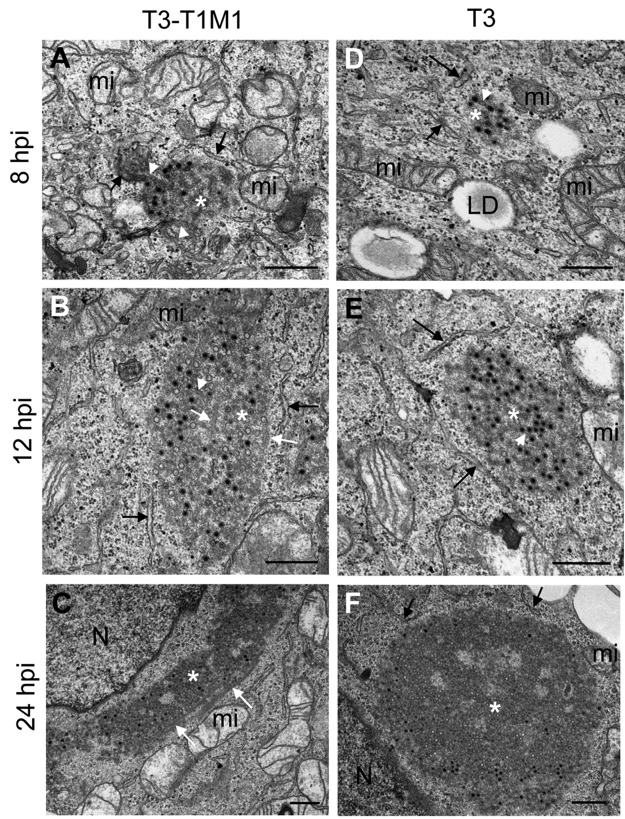
Most viruses that replicate in the cytoplasm of host cells form neo-organelles that serve as sites of viral genome replication and particle assembly. These highly specialized structures concentrate viral replication proteins and nucleic acids, prevent the activation of cell-intrinsic defenses, and coordinate the release of progeny particles. Despite the importance of inclusion complexes in viral replication, there are key gaps in the knowledge of how these organelles form and mediate their functions. Reoviruses are nonenveloped, double-stranded RNA (dsRNA) viruses that serve as tractable experimental models for studies of dsRNA virus replication and pathogenesis. Following reovirus entry into cells, replication occurs in large cytoplasmic structures termed inclusions that fill with progeny virions. Reovirus inclusions are nucleated by viral nonstructural proteins, which in turn recruit viral structural proteins for genome replication and particle assembly. Components of reovirus inclusions are poorly understood, but these structures are generally thought to be devoid of membranes. We used transmission electron microscopy and three-dimensional image reconstructions to visualize reovirus inclusions in infected cells. These studies revealed that reovirus inclusions form within a membranous network. Viral inclusions contain filled and empty viral particles and microtubules and appose mitochondria and rough endoplasmic reticulum (RER). Immunofluorescence confocal microscopy analysis demonstrated that markers of the ER and ER-Golgi intermediate compartment (ERGIC) codistribute with inclusions during infection, as does dsRNA. dsRNA colocalizes with the viral protein σNS and an ERGIC marker inside inclusions. These findings suggest that cell membranes within reovirus inclusions form a scaffold to coordinate viral replication and assembly.
Viruses alter the architecture of host cells to form an intracellular environment conducive to viral replication. This step in viral infection requires the concerted action of viral and host components and is potentially vulnerable to pharmacological intervention. Reoviruses form large cytoplasmic replication sites called inclusions, which have been described as membrane-free structures. Despite the importance of inclusions in the reovirus replication cycle, little is known about their formation and composition. We used light and electron microscopy to demonstrate that reovirus inclusions are membrane-containing structures and that the endoplasmic reticulum (ER) and the ER-Golgi intermediate compartment interact closely with these viral organelles. These findings enhance our understanding of the cellular machinery usurped by viruses to form inclusion organelles and complete an infectious cycle. This information, in turn, may foster the development of antiviral drugs that impede this essential viral replication step.
大多数在宿主细胞质中复制的病毒会形成新细胞器,作为病毒基因组复制和颗粒组装的场所。这些高度特化的结构聚集病毒复制蛋白和核酸,防止细胞固有防御的激活,并协调子代颗粒的释放。尽管包涵体复合物在病毒复制中很重要,但关于这些细胞器如何形成及其功能介导机制的知识仍存在关键空白。呼肠孤病毒是无包膜的双链RNA(dsRNA)病毒,是研究dsRNA病毒复制和发病机制的易于处理的实验模型。呼肠孤病毒进入细胞后,在称为包涵体的大型细胞质结构中进行复制,包涵体中充满子代病毒粒子。呼肠孤病毒包涵体由病毒非结构蛋白成核,这些非结构蛋白进而招募病毒结构蛋白进行基因组复制和颗粒组装。呼肠孤病毒包涵体的组成成分了解甚少,但这些结构通常被认为没有膜。我们使用透射电子显微镜和三维图像重建技术来观察感染细胞中的呼肠孤病毒包涵体。这些研究表明,呼肠孤病毒包涵体在膜性网络内形成。病毒包涵体包含充满和空的病毒粒子以及微管,并与线粒体和粗面内质网(RER)相邻。免疫荧光共聚焦显微镜分析表明,内质网(ER)和内质网-高尔基体中间区室(ERGIC)的标记物在感染期间与包涵体共分布,dsRNA也是如此。dsRNA与病毒蛋白σNS和包涵体内的ERGIC标记物共定位。这些发现表明,呼肠孤病毒包涵体内的细胞膜形成了一个支架,以协调病毒的复制和组装。
病毒改变宿主细胞的结构,以形成有利于病毒复制的细胞内环境。病毒感染的这一步骤需要病毒和宿主成分的协同作用,并且可能容易受到药物干预。呼肠孤病毒形成称为包涵体的大型细胞质复制位点,这些位点被描述为无膜结构。尽管包涵体在呼肠孤病毒复制周期中很重要,但对其形成和组成知之甚少。我们使用光学和电子显微镜证明,呼肠孤病毒包涵体是含有膜的结构,内质网(ER)和内质网-高尔基体中间区室与这些病毒细胞器密切相互作用。这些发现增强了我们对病毒篡夺细胞机制以形成包涵体细胞器并完成感染周期的理解。反过来,这些信息可能会促进开发阻碍这一关键病毒复制步骤的抗病毒药物。