Department of Medical Sciences, University of Torino, Torino, Italy.
Translational Center for Regenerative Medicine, University of Torino, Torino, Italy.
Int J Mol Med. 2014 May;33(5):1055-63. doi: 10.3892/ijmm.2014.1663. Epub 2014 Feb 20.
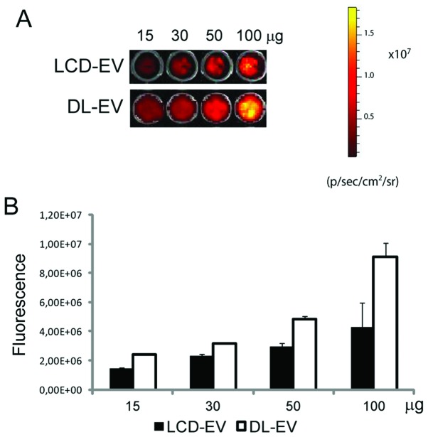
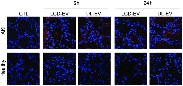
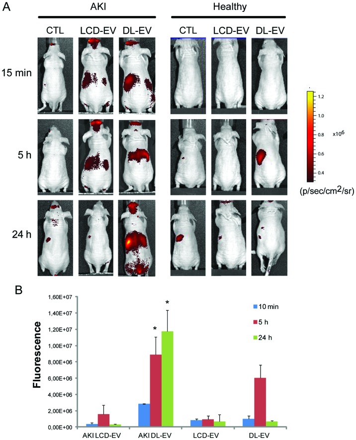
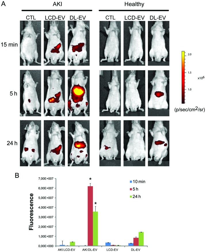
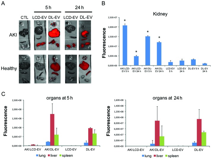
Mesenchymal stem cells (MSCs) contribute to the recovery of tissue injury, providing a paracrine support. Cell-derived extracellular vesicles (EVs), carrying membrane and cytoplasmatic constituents of the cell of origin, have been described as a fundamental mechanism of intercellular communication. We previously demonstrated that EVs derived from human MSCs accelerated recovery following acute kidney injury (AKI) in vivo. The aim of the present study was to investigate the biodistribution and the renal localization of EVs in AKI. For this purpose, two methods for EV labeling suitable for in vivo tracking with optical imaging (OI), were employed using near infrared (NIR) dye (DiD): i) labeled EVs were generated by MSCs pre-incubated with NIR dye and collected from cell supernatants; ii) purified EVs were directly labeled with NIR dye. EVs obtained with these two procedures were injected intravenously (i.v.) into mice with glycerol-induced AKI and into healthy mice to compare the efficacy of the two labeling methods for in vivo detection of EVs at the site of damage. We found that the labeled EVs accumulated specifically in the kidneys of the mice with AKI compared with the healthy controls. After 5 h, the EVs were detectable in whole body images and in dissected kidneys by OI with both types of labeling procedures. The directly labeled EVs showed a higher and brighter fluorescence compared with the labeled EVs produced by cells. The signal generated by the directly labeled EVs was maintained in time, but provided a higher background than that of the labeled EVs produced by cells. The comparison of the two methods indicated that the latter displayed a greater specificity for the injured kidney.
间充质干细胞 (MSCs) 通过旁分泌支持作用促进组织损伤的恢复。细胞衍生的细胞外囊泡 (EVs) 携带起源细胞的膜和细胞质成分,被认为是细胞间通讯的基本机制。我们之前证明,源自人 MSCs 的 EVs 可加速体内急性肾损伤 (AKI) 的恢复。本研究旨在研究 EV 在 AKI 中的体内分布和肾脏定位。为此,使用近红外 (NIR) 染料 (DiD) 采用两种适合光学成像 (OI) 体内示踪的 EV 标记方法:i)通过预先用 NIR 染料孵育的 MSC 生成标记的 EV,并从细胞上清液中收集;ii)直接用 NIR 染料标记纯化的 EV。用这两种方法获得的 EV 被静脉内 (i.v.) 注射到甘油诱导的 AKI 小鼠和健康小鼠中,以比较两种标记方法在损伤部位体内检测 EV 的效果。我们发现,与健康对照组相比,标记的 EV 特异性地积聚在 AKI 小鼠的肾脏中。5 小时后,通过 OI 可以在全身图像和解剖的肾脏中检测到两种类型的标记程序的标记 EV。与由细胞产生的标记 EV 相比,直接标记的 EV 显示出更高和更亮的荧光。直接标记的 EV 产生的信号随时间保持,但与由细胞产生的标记 EV 相比,其背景更高。两种方法的比较表明,后者对受损肾脏具有更高的特异性。