Garrovo Chiara, Bergamin Natascha, Bates Dave, Cesselli Daniela, Beltrami Antonio Paolo, Lorenzon Andrea, Ferrari Roberto, Alberto Beltrami Carlo, Lorusso Vito, Biffi Stefania
Optical Imaging Laboratory, Cluster in Biomedicine (CBM scrl), 34149 Trieste, Italy.
Int J Mol Imaging. 2013;2013:426961. doi: 10.1155/2013/426961. Epub 2013 Jan 17.
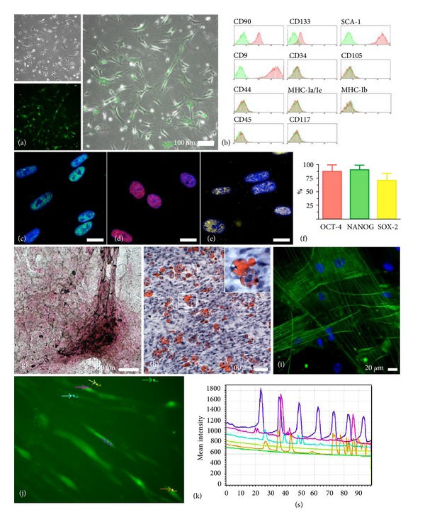
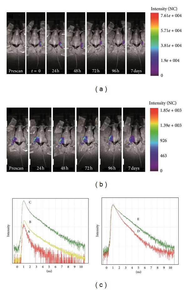
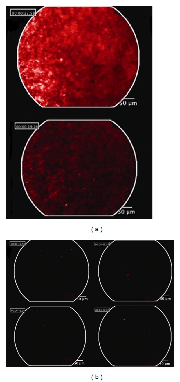
Stem cells are characterized by the ability to renew themselves and to differentiate into specialized cell types, while stem cell therapy is believed to treat a number of different human diseases through either cell regeneration or paracrine effects. Herein, an in vivo and ex vivo near infrared time domain (NIR TD) optical imaging study was undertaken to evaluate the migratory ability of murine adipose tissue-derived multipotent adult stem cells [mAT-MASC] after intramuscular injection in mice. In vivo NIR TD optical imaging data analysis showed a migration of DiD-labelled mAT-MASC in the leg opposite the injection site, which was confirmed by a fibered confocal microendoscopy system. Ex vivo NIR TD optical imaging results showed a systemic distribution of labelled cells. Considering a potential microenvironmental contamination, a cross-validation study by multimodality approaches was followed: mAT-MASC were isolated from male mice expressing constitutively eGFP, which was detectable using techniques of immunofluorescence and qPCR. Y-chromosome positive cells, injected into wild-type female recipients, were detected by FISH. Cross-validation confirmed the data obtained by in vivo/ex vivo TD optical imaging analysis. In summary, our data demonstrates the usefulness of NIR TD optical imaging in tracking delivered cells, giving insights into the migratory properties of the injected cells.
干细胞的特点是能够自我更新并分化为特定的细胞类型,而干细胞疗法被认为可以通过细胞再生或旁分泌效应来治疗多种不同的人类疾病。在此,我们进行了一项体内和体外近红外时域(NIR TD)光学成像研究,以评估小鼠脂肪组织来源的多能成体干细胞[mAT-MASC]在小鼠肌肉注射后的迁移能力。体内NIR TD光学成像数据分析显示,DiD标记的mAT-MASC在注射部位对侧的腿部发生了迁移,这一点通过纤维共聚焦显微内镜系统得到了证实。体外NIR TD光学成像结果显示标记细胞呈全身分布。考虑到潜在的微环境污染,我们采用多模态方法进行了交叉验证研究:从组成性表达eGFP的雄性小鼠中分离出mAT-MASC,可使用免疫荧光和qPCR技术检测到eGFP。通过FISH检测注射到野生型雌性受体中的Y染色体阳性细胞。交叉验证证实了体内/体外TD光学成像分析获得的数据。总之,我们的数据证明了NIR TD光学成像在追踪递送细胞方面的有用性,有助于深入了解注射细胞的迁移特性。