Francischetti Ivo M B, Gordon Emile, Bizzarro Bruna, Gera Nidhi, Andrade Bruno B, Oliveira Fabiano, Ma Dongying, Assumpção Teresa C F, Ribeiro José M C, Pena Mirna, Qi Chen-Feng, Diouf Ababacar, Moretz Samuel E, Long Carole A, Ackerman Hans C, Pierce Susan K, Sá-Nunes Anderson, Waisberg Michael
Laboratory of Malaria and Vector Research, National Institute of Allergy and Infectious Diseases, National Institutes of Health, Rockville, Maryland, United States of America.
Laboratory of Immunogenetics, National Institute of Allergy and Infectious Diseases, National Institutes of Health, Rockville, Maryland, United States of America.
PLoS One. 2014 Feb 28;9(2):e87140. doi: 10.1371/journal.pone.0087140. eCollection 2014.
The role of intracellular radical oxygen species (ROS) in pathogenesis of cerebral malaria (CM) remains incompletely understood.
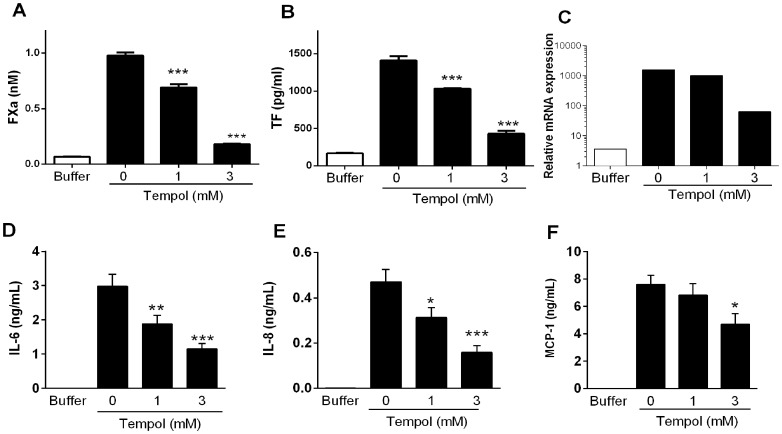
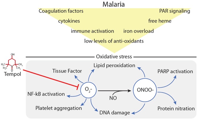
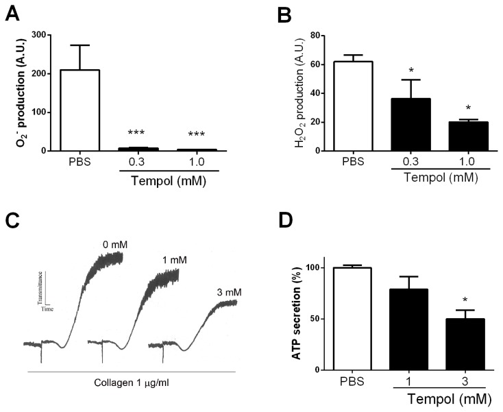
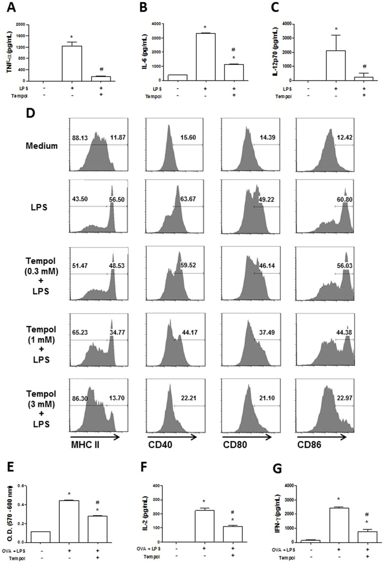
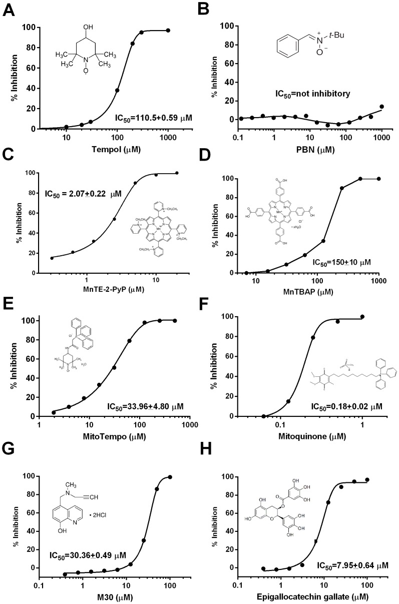
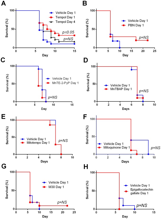
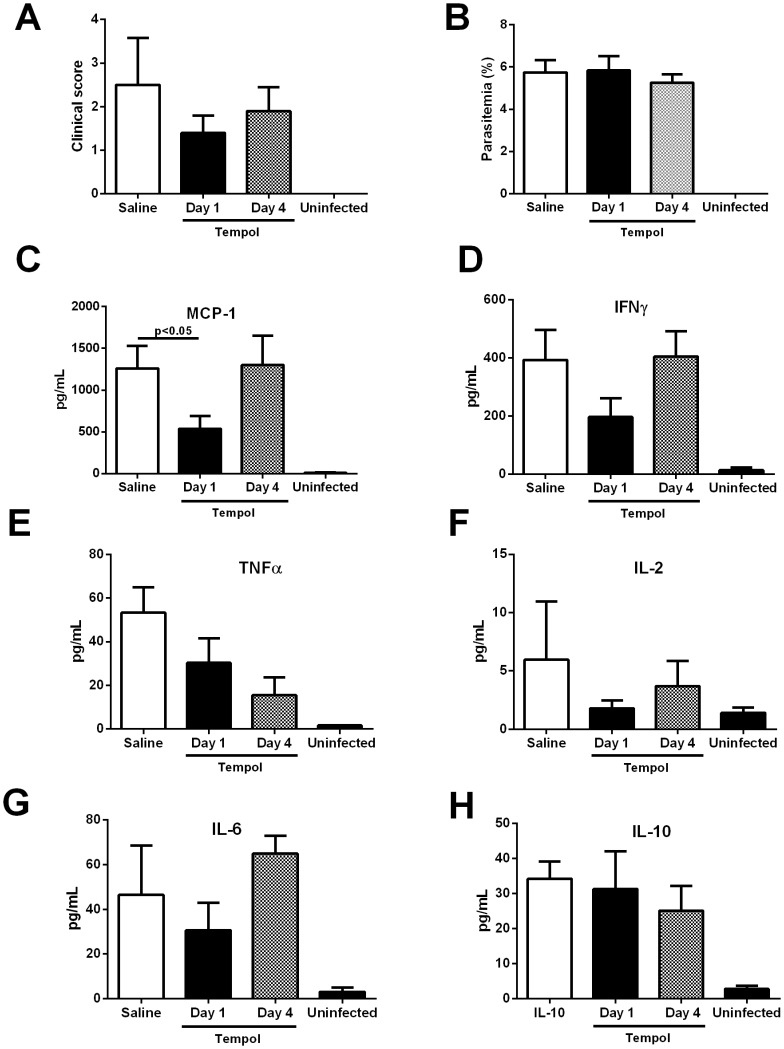
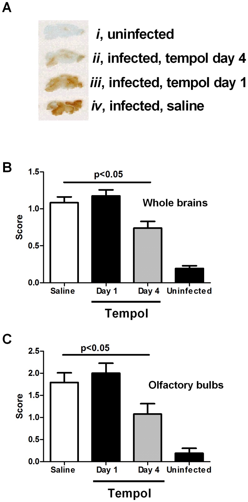
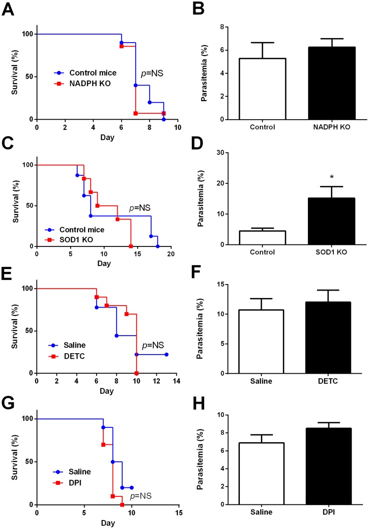
We undertook testing Tempol--a superoxide dismutase (SOD) mimetic and pleiotropic intracellular antioxidant--in cells relevant to malaria pathogenesis in the context of coagulation and inflammation. Tempol was also tested in a murine model of CM induced by Plasmodium berghei Anka infection. Tempol was found to prevent transcription and functional expression of procoagulant tissue factor in endothelial cells (ECs) stimulated by lipopolysaccharide (LPS). This effect was accompanied by inhibition of IL-6, IL-8, and monocyte chemoattractant protein (MCP-1) production. Tempol also attenuated platelet aggregation and human promyelocytic leukemia HL60 cells oxidative burst. In dendritic cells, Tempol inhibited LPS-induced production of TNF-α, IL-6, and IL-12p70, downregulated expression of co-stimulatory molecules, and prevented antigen-dependent lymphocyte proliferation. Notably, Tempol (20 mg/kg) partially increased the survival of mice with CM. Mechanistically, treated mice had lowered plasma levels of MCP-1, suggesting that Tempol downmodulates EC function and vascular inflammation. Tempol also diminished blood brain barrier permeability associated with CM when started at day 4 post infection but not at day 1, suggesting that ROS production is tightly regulated. Other antioxidants-such as α-phenyl N-tertiary-butyl nitrone (PBN; a spin trap), MnTe-2-PyP and MnTBAP (Mn-phorphyrin), Mitoquinone (MitoQ) and Mitotempo (mitochondrial antioxidants), M30 (an iron chelator), and epigallocatechin gallate (EGCG; polyphenol from green tea) did not improve survival. By contrast, these compounds (except PBN) inhibited Plasmodium falciparum growth in culture with different IC50s. Knockout mice for SOD1 or phagocyte nicotinamide adenine dinucleotide phosphate (NADPH) oxidase (gp91(phox-/-)) or mice treated with inhibitors of SOD (diethyldithiocarbamate) or NADPH oxidase (diphenyleneiodonium) did not show protection or exacerbation for CM.
Results with Tempol suggest that intracellular ROS contribute, in part, to CM pathogenesis. Therapeutic targeting of intracellular ROS in CM is discussed.
细胞内活性氧(ROS)在脑型疟疾(CM)发病机制中的作用仍未完全明确。
我们在与疟疾发病机制相关的凝血和炎症细胞中,对替莫泊尔(一种超氧化物歧化酶(SOD)模拟物和多效性细胞内抗氧化剂)进行了测试。还在伯氏疟原虫安氏株感染诱导的CM小鼠模型中对替莫泊尔进行了测试。发现替莫泊尔可预防脂多糖(LPS)刺激的内皮细胞(ECs)中促凝血组织因子的转录和功能表达。这一效应伴随着白细胞介素-6(IL-6)、白细胞介素-8(IL-8)和单核细胞趋化蛋白(MCP-1)产生的抑制。替莫泊尔还减弱了血小板聚集和人早幼粒细胞白血病HL60细胞的氧化爆发。在树突状细胞中,替莫泊尔抑制LPS诱导的肿瘤坏死因子-α(TNF-α)、IL-6和IL-12p70的产生,下调共刺激分子的表达,并阻止抗原依赖性淋巴细胞增殖。值得注意的是,替莫泊尔(20mg/kg)可部分提高CM小鼠的存活率。从机制上讲,经治疗的小鼠血浆中MCP-1水平降低,表明替莫泊尔下调了EC功能和血管炎症。在感染后第4天开始使用替莫泊尔可降低与CM相关的血脑屏障通透性,但在第1天开始使用则无效,这表明ROS的产生受到严格调控。其他抗氧化剂,如α-苯基N-叔丁基硝酮(PBN;一种自旋捕获剂)、MnTe-2-PyP和MnTBAP(锰卟啉)、米托醌(MitoQ)和米托替莫(线粒体抗氧化剂)、M30(一种铁螯合剂)以及表没食子儿茶素没食子酸酯(EGCG;绿茶中的多酚)均未改善存活率。相比之下,这些化合物(除PBN外)在不同半数抑制浓度(IC50)下抑制了恶性疟原虫在培养物中的生长。超氧化物歧化酶1(SOD1)基因敲除小鼠、吞噬细胞烟酰胺腺嘌呤二核苷酸磷酸(NADPH)氧化酶(gp91(phox-/-))基因敲除小鼠或用SOD抑制剂(二乙基二硫代氨基甲酸盐)或NADPH氧化酶抑制剂(二苯基碘鎓)处理的小鼠对CM均未表现出保护作用或病情加重。
替莫泊尔的实验结果表明,细胞内ROS在一定程度上参与了CM的发病机制。文中讨论了针对CM中细胞内ROS的治疗靶点。