European Synchrotron Radiation Facility, Grenoble, France ; Berlin-Brandenburg School for Regenerative Therapies & Julius Wolff Institut, Charité, Universitätsmedizin Berlin, Germany.
European Synchrotron Radiation Facility, Grenoble, France ; Université de Lyon, CREATIS, CNRS UMR5220, INSA-Lyon, Lyon, France.
PLoS One. 2014 Feb 21;9(2):e88481. doi: 10.1371/journal.pone.0088481. eCollection 2014.
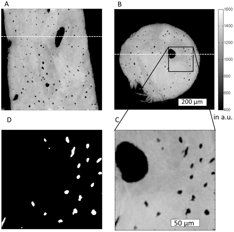
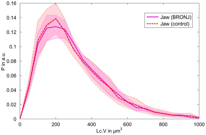
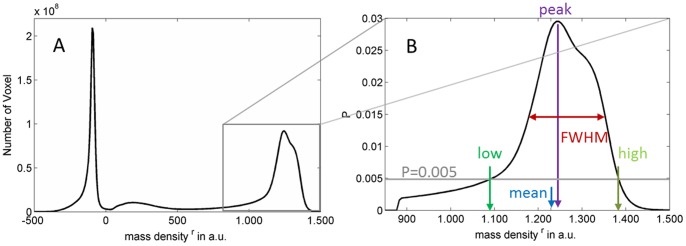
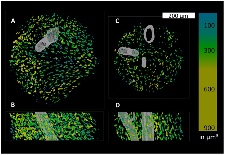
Osteonecrosis of the jaw, in association with bisphosphonates (BRONJ) used for treating osteoporosis or cancer, is a severe and most often irreversible side effect whose underlying pathophysiological mechanisms remain largely unknown. Osteocytes are involved in bone remodeling and mineralization where they orchestrate the delicate equilibrium between osteoclast and osteoblast activity and through the active process called osteocytic osteolysis. Here, we hypothesized that (i) changes of the mineralized tissue matrix play a substantial role in the pathogenesis of BRONJ, and (ii) the osteocyte lacunar morphology is altered in BRONJ. Synchrotron µCT with phase contrast is an appropriate tool for assessing both the 3D morphology of the osteocyte lacunae and the bone matrix mass density. Here, we used this technique to investigate the mass density distribution and 3D osteocyte lacunar properties at the sub-micrometer scale in human bone samples from the jaw, femur and tibia. First, we compared healthy human jaw bone to human tibia and femur in order to assess the specific differences and address potential explanations of why the jaw bone is exclusively targeted by the necrosis as a side effect of BP treatment. Second, we investigated the differences between BRONJ and control jaw bone samples to detect potential differences which could aid an improved understanding of the course of BRONJ. We found that the apparent mass density of jaw bone was significantly smaller compared to that of tibia, consistent with a higher bone turnover in the jaw bone. The variance of the lacunar volume distribution was significantly different depending on the anatomical site. The comparison between BRONJ and control jaw specimens revealed no significant increase in mineralization after BP. We found a significant decrease in osteocyte-lacunar density in the BRONJ group compared to the control jaw. Interestingly, the osteocyte-lacunar volume distribution was not altered after BP treatment.
颌骨骨坏死(BRONJ)与双膦酸盐(BRONJ)有关,后者用于治疗骨质疏松症或癌症,是一种严重且通常不可逆转的副作用,其潜在的病理生理机制在很大程度上尚不清楚。成骨细胞参与骨重塑和矿化,在那里它们协调破骨细胞和成骨细胞活性之间的微妙平衡,并通过称为成骨细胞性骨溶解的主动过程。在这里,我们假设(i)矿化组织基质的变化在 BRONJ 的发病机制中起重要作用,(ii)BRONJ 中的成骨细胞陷窝形态发生改变。同步加速器μCT 与相位对比是评估成骨细胞陷窝的 3D 形态和骨基质质量密度的适当工具。在这里,我们使用该技术在颌骨、股骨和胫骨的人骨样本中研究了亚微米尺度的骨基质质量密度分布和 3D 成骨细胞陷窝特性。首先,我们比较了健康人的颌骨与人类胫骨和股骨,以评估特定的差异,并为颌骨为何仅作为 BP 治疗的副作用而成为坏死的靶向提供潜在的解释。其次,我们研究了 BRONJ 和对照颌骨样本之间的差异,以检测潜在的差异,这可能有助于更好地理解 BRONJ 的发病过程。我们发现,与胫骨相比,颌骨的表观质量密度明显较小,这与颌骨的更高骨转换一致。陷窝体积分布的方差根据解剖部位而有显著差异。BRONJ 和对照颌骨标本之间的比较显示,BP 后矿物质化没有明显增加。与对照组相比,BRONJ 组的成骨细胞陷窝密度显着降低。有趣的是,BP 治疗后成骨细胞陷窝体积分布没有改变。