Aab Cardiovascular Research Institute and the Department of Medicine, University of Rochester Medical Center, Rochester, New York, United States of America ; Orthopaedic Department, The First Affiliated Hospital of Nanjing Medical University, Jiangsu, China.
Center for Musculoskeletal Research and the Department of Orthopaedics and Rehabilitation, University of Rochester Medical Center, Rochester, New York, United States of America.
PLoS One. 2014 Feb 19;9(2):e89127. doi: 10.1371/journal.pone.0089127. eCollection 2014.
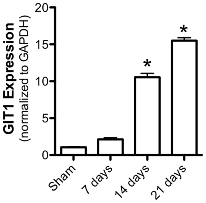
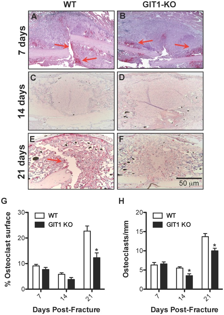
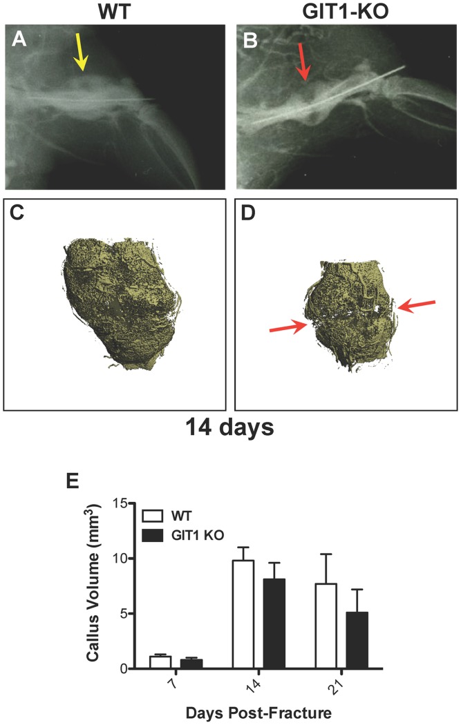
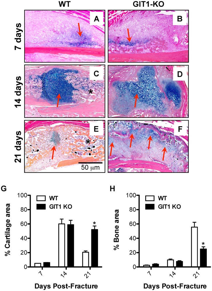
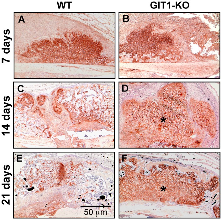
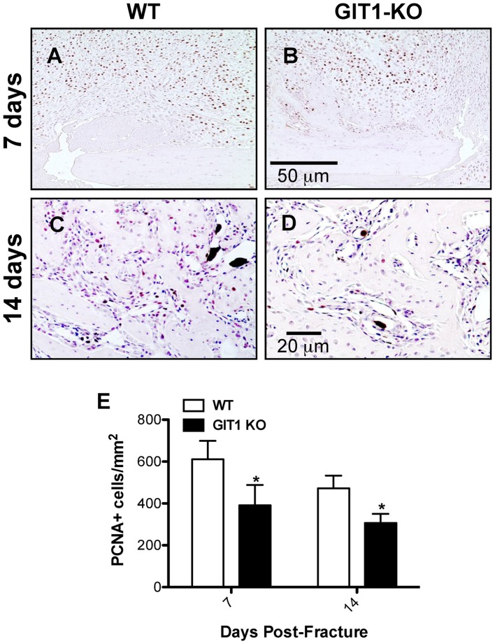
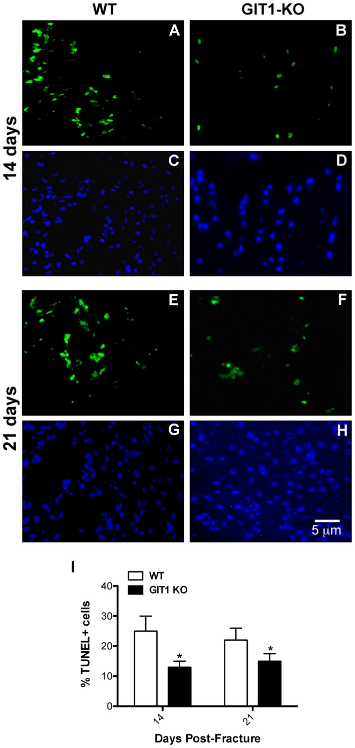
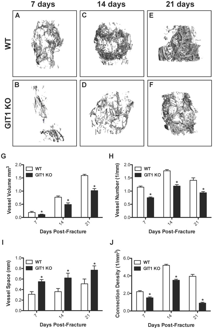
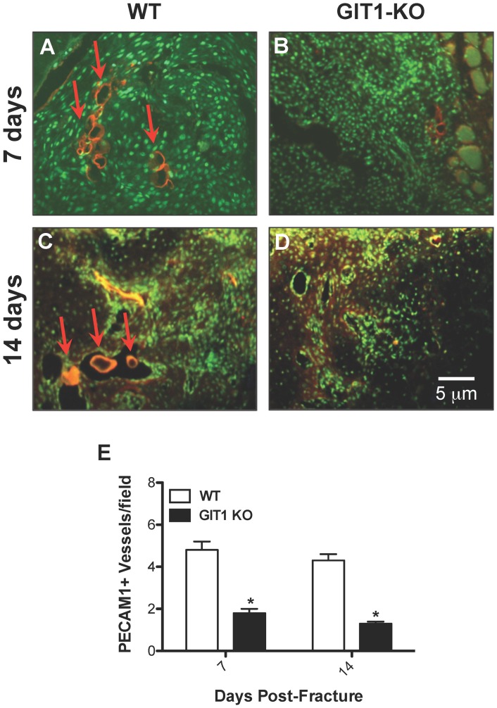
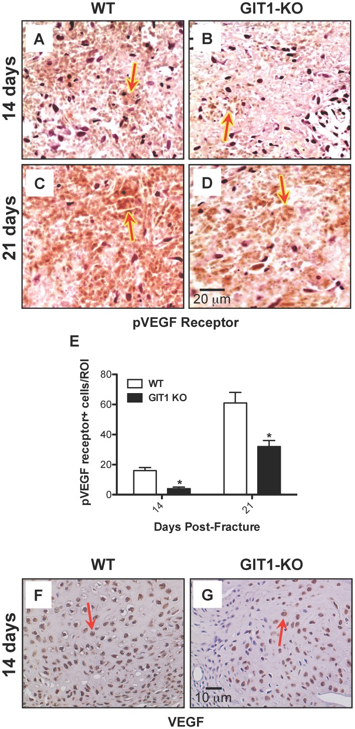
G protein coupled receptor kinase 2 (GRK2) interacting protein-1 (GIT1), is a scaffold protein that plays an important role in angiogenesis and osteoclast activity. We have previously demonstrated that GIT1 knockout (GIT1 KO) mice have impaired angiogenesis and dysregulated osteoclast podosome formation leading to a reduction in the bone resorbing ability of these cells. Since both angiogenesis and osteoclast-mediated bone remodeling are involved in the fracture healing process, we hypothesized that GIT1 participates in the normal progression of repair following bone injury. In the present study, comparison of fracture healing in wild type (WT) and GIT1 KO mice revealed altered healing in mice with loss of GIT1 function. Alcian blue staining of fracture callus indicated a persistence of cartilagenous matrix in day 21 callus samples from GIT1 KO mice which was temporally correlated with increased type 2 collagen immunostaining. GIT1 KO mice also showed a decrease in chondrocyte proliferation and apoptosis at days 7 and 14, as determined by PCNA and TUNEL staining. Vascular microcomputed tomography analysis of callus samples at days 7, 14 and 21 revealed decreased blood vessel volume, number, and connection density in GIT1 KO mice compared to WT controls. Correlating with this, VEGF-A, phospho-VEGFR2 and PECAM1 (CD31) were decreased in GIT1 KO mice, indicating reduced angiogenesis with loss of GIT1. Finally, calluses from GIT1 KO mice displayed a reduced number of tartrate resistant acid phosphatase-positive osteoclasts at days 14 and 21. Collectively, these results indicate that GIT1 is an important signaling participant in fracture healing, with gene ablation leading to reduced callus vascularity and reduced osteoclast number in the healing callus.
G 蛋白偶联受体激酶 2(GRK2)相互作用蛋白-1(GIT1)是一种支架蛋白,在血管生成和破骨细胞活性中发挥重要作用。我们之前已经证明,GIT1 敲除(GIT1 KO)小鼠的血管生成受损,破骨细胞足突形成失调,导致这些细胞的骨吸收能力降低。由于血管生成和破骨细胞介导的骨重塑都参与骨折愈合过程,我们假设 GIT1 参与骨损伤后的正常修复过程。在本研究中,比较野生型(WT)和 GIT1 KO 小鼠的骨折愈合情况,发现 GIT1 功能丧失的小鼠的愈合情况发生了改变。骨折痂内的阿尔辛蓝染色表明,GIT1 KO 小鼠的第 21 天骨折痂样本中软骨基质持续存在,这与 2 型胶原免疫染色的增加时间相关。GIT1 KO 小鼠在第 7 天和第 14 天的软骨细胞增殖和凋亡也减少,这通过 PCNA 和 TUNEL 染色来确定。第 7、14 和 21 天的骨痂样本的血管微计算机断层扫描分析显示,与 WT 对照组相比,GIT1 KO 小鼠的血管体积、数量和连接密度减少。与此相关的是,GIT1 KO 小鼠的 VEGF-A、磷酸化 VEGFR2 和 PECAM1(CD31)减少,表明 GIT1 缺失导致血管生成减少。最后,GIT1 KO 小鼠的骨痂中抗酒石酸酸性磷酸酶阳性破骨细胞的数量在第 14 天和第 21 天减少。总的来说,这些结果表明 GIT1 是骨折愈合中一个重要的信号参与者,基因缺失导致愈合骨痂中的骨痂血管减少和破骨细胞数量减少。