Chen Peng, Gu Wan-Li, Gong Ming-Zhi, Wang Jun, Li Dong-Qing
Department of Trauma Orthopedics, The Second Hospital of Shandong University, Jinan, 250033, People's Republic of China.
Department of Operating Theater, The Second Hospital of Shandong University, No. 247, Beiyuan Street, Jinan, 250033, Shandong Province, People's Republic of China.
BMC Musculoskelet Disord. 2017 Jul 28;18(1):320. doi: 10.1186/s12891-017-1653-7.
Although tibial plateau fracture is an uncommon injury, its regulation is challenging and there are some influencing factors, including the effects of severe bone displacement, depression and cancellous bone cartilage, and inevitable cartilage damage. And GIT1 plays an important role in bone mass and 78 osteoblast cell migration.
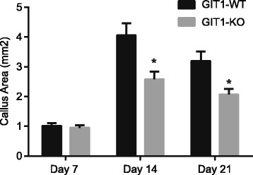
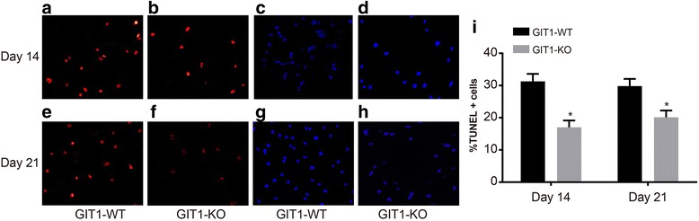
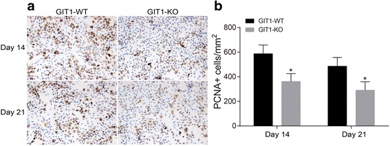
The study used 72 C57/BL6 mice. A tibial plateau fracture model was established by using mice with the same number of GIT1 gene deletions (the experimental group) and their wild-type littermates (the control group). Joint and bone callus recovery were evaluated by X-ray and CT thin layer scans. Micro CT assay and histomorphometry were conducted in order to evaluate the volume of newly formed blood vessels. Type II collagen expression in tibial tissues after tibial plateau fracture were detected by immunohistochemistry after 7, 14 and 21 days. The number of proliferating cell nuclear antigen (PCNA) positive cells after tibial plateau fracture was tested by immunohistochemistry after 14 and 21 days. The terminal deoxynucleotidyl transferase-mediated dUTP-biotin nick end labeling (TUNEL) staining was conducted after 14 and 21 days in order to test chondrocyte apoptosis in tibial tissues after tibial plateau fracture.
The GIT1 gene deletion group mice spent less time on the rotating rod than the control group mice (P < 0.05). Compared with the control group, postoperative recovery was retarded, because GIT1 gene deletion slowed down neovascularization after tibial plateau fracture (P < 0.05). Compared with the control group, mouse type II collagen expression significantly decreased in the GIT1 gene deletion group, and the proportion of PCNA positive cells significantly decreased (P < 0.05). The TUNEL results indicate that GIT1 gene deletion led to reduced chondrocyte apoptosis.
GIT1 gene deletion can inhibit chondrocyte proliferation and apoptosis during the recovery of tibial plateau fracture, so as to delay chondrocyte differentiation and tibial plateau fracture healing.
虽然胫骨平台骨折是一种不常见的损伤,但其治疗具有挑战性,存在一些影响因素,包括严重的骨移位、凹陷和松质骨软骨的影响以及不可避免的软骨损伤。并且GIT1在骨量和78个成骨细胞迁移中起重要作用。
本研究使用72只C57/BL6小鼠。通过使用相同数量的GIT1基因缺失小鼠(实验组)及其野生型同窝小鼠(对照组)建立胫骨平台骨折模型。通过X射线和CT薄层扫描评估关节和骨痂恢复情况。进行显微CT分析和组织形态计量学以评估新形成血管的体积。在胫骨平台骨折后7、14和21天,通过免疫组织化学检测胫骨组织中II型胶原蛋白的表达。在胫骨平台骨折后14和21天,通过免疫组织化学检测增殖细胞核抗原(PCNA)阳性细胞的数量。在胫骨平台骨折后14和21天进行末端脱氧核苷酸转移酶介导的dUTP-生物素缺口末端标记(TUNEL)染色,以检测胫骨组织中的软骨细胞凋亡。
GIT1基因缺失组小鼠在转棒上花费的时间比对照组小鼠少(P < 0.05)。与对照组相比,术后恢复延迟,因为GIT1基因缺失减缓了胫骨平台骨折后的新生血管形成(P < 0.05)。与对照组相比,GIT1基因缺失组小鼠II型胶原蛋白表达明显降低,PCNA阳性细胞比例明显降低(P < 0.05)。TUNEL结果表明,GIT1基因缺失导致软骨细胞凋亡减少。
GIT1基因缺失可抑制胫骨平台骨折恢复过程中软骨细胞的增殖和凋亡,从而延缓软骨细胞分化和胫骨平台骨折愈合。