• 文献检索
  • 文档翻译
  • 深度研究
  • 学术资讯
  • Suppr Zotero 插件Zotero 插件
  • 邀请有礼
  • 套餐&价格
  • 历史记录
应用&插件
Suppr Zotero 插件Zotero 插件浏览器插件Mac 客户端Windows 客户端微信小程序
定价
高级版会员购买积分包购买API积分包
服务
文献检索文档翻译深度研究API 文档MCP 服务
关于我们
关于 Suppr公司介绍联系我们用户协议隐私条款
关注我们

Suppr 超能文献

核心技术专利:CN118964589B侵权必究
粤ICP备2023148730 号-1Suppr @ 2026

文献检索

告别复杂PubMed语法,用中文像聊天一样搜索,搜遍4000万医学文献。AI智能推荐,让科研检索更轻松。

立即免费搜索

文件翻译

保留排版,准确专业,支持PDF/Word/PPT等文件格式,支持 12+语言互译。

免费翻译文档

深度研究

AI帮你快速写综述,25分钟生成高质量综述,智能提取关键信息,辅助科研写作。

立即免费体验

“活化石”双壳贝类 Neotrigonia 中几丁质层的早期生物矿化作用

Early stage biomineralization in the periostracum of the 'living fossil' bivalve Neotrigonia.

机构信息

Departamento de Estratigrafía y Paleontología, Facultad de Ciencias, Universidad de Granada, Granada, Spain.

Departamento de Biología Animal, Facultad de Ciencias, Universidad de Málaga, Málaga, Spain.

出版信息

PLoS One. 2014 Feb 25;9(2):e90033. doi: 10.1371/journal.pone.0090033. eCollection 2014.

DOI:10.1371/journal.pone.0090033
PMID:24587202
原文链接:https://pmc.ncbi.nlm.nih.gov/articles/PMC3934977/
Abstract

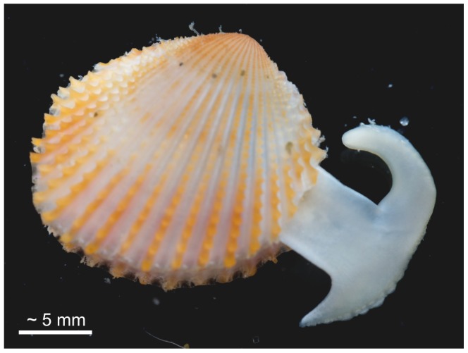
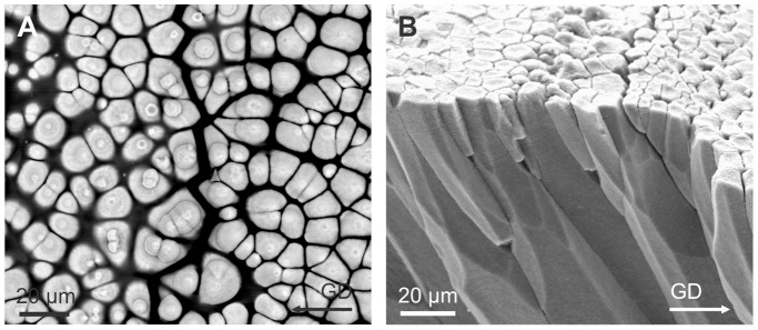
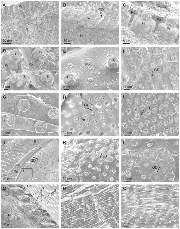
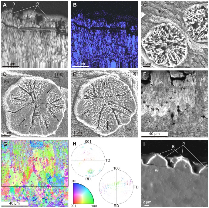
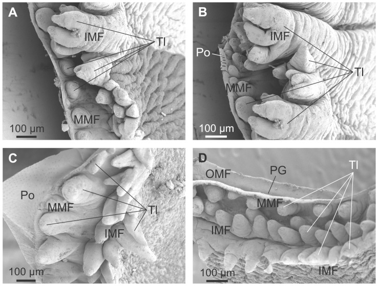
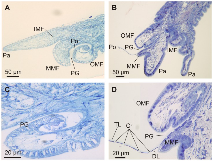
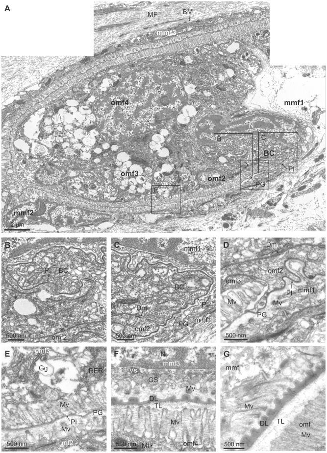
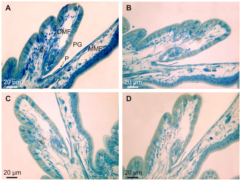
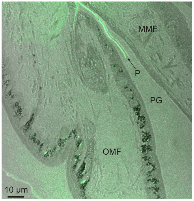
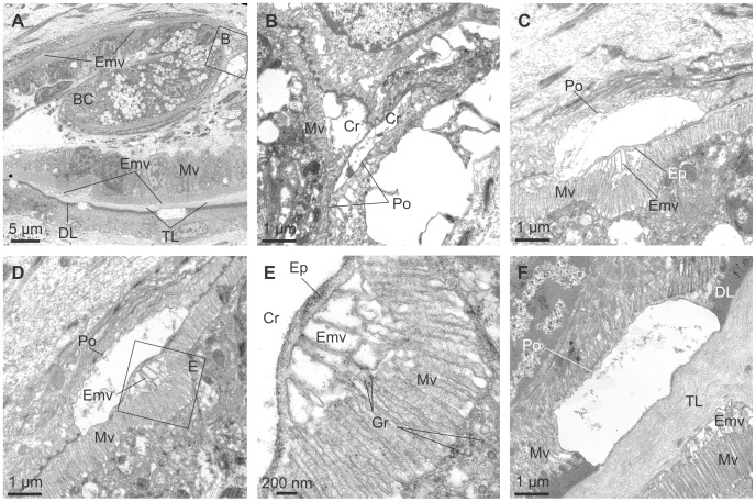
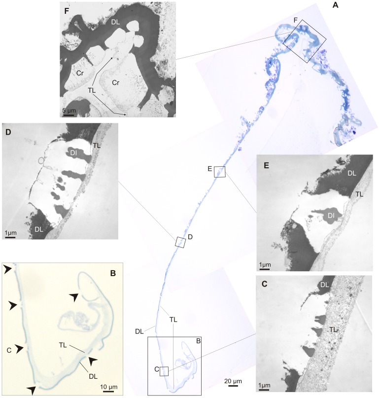
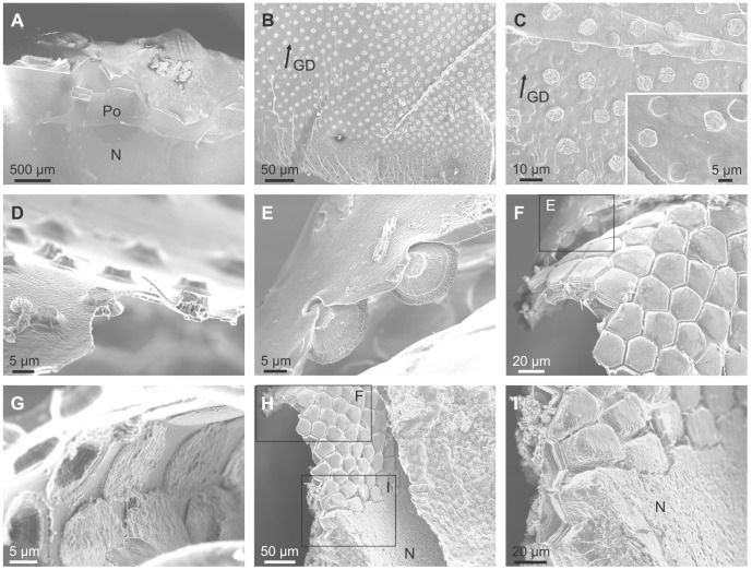
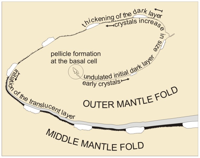
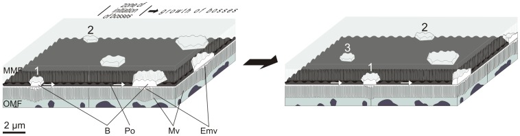
A detailed investigation of the shell formation of the palaeoheterodont 'living fossil' Neotrigonia concentrated on the timing and manufacture of the calcified 'bosses' which stud the outside of all trigonioid bivalves (extant and fossil) has been conducted. Electron microscopy and optical microscopy revealed that Neotrigonia spp. have a spiral-shaped periostracal groove. The periostracum itself is secreted by the basal cell, as a thin dark pellicle, becoming progressively transformed into a thin dark layer by additions of secretions from the internal outer mantle fold. Later, intense secretion of the internal surface of the outer mantle fold forms a translucent layer, which becomes transformed by tanning into a dark layer. The initiation of calcified bosses occurred at a very early stage of periostracum formation, deep within the periostracal groove immediately below the initialmost dark layer. At this stage, they consist of a series of polycyclically twinned crystals. The bosses grow as the periostracum traverse through the periostracal groove, in coordination with the thickening of the dark periostracal layer and until, upon reaching the mantle edge, they impinge upon each other and become transformed into large prisms separated by dark periostracal walls. In conclusion, the initial bosses and the external part of the prismatic layer are fully intraperiostracal. With later growth, the prisms transform into fibrous aggregates, although the details of the process are unknown. This reinforces the relationships with other groups that have the ability to form intraperiostracal calcifications, for example the unionoids with which the trigonioids form the clade Paleoheterodonta. The presence of similar structures in anomalodesmatans and other euheterodonts raises the question of whether this indicates a relationship or represents a convergence. The identification of very early calcification within an organic sheet has interesting implications for our understanding of how shells may have evolved.

摘要

对古有齿蛤“活化石” Neo-trigonia 的壳形成进行了详细研究,重点关注了覆盖所有三角蛤(现存和化石)外部的钙化“凸瘤”的形成时间和制造过程。电子显微镜和光学显微镜显示,Neotrigonia spp. 具有螺旋状的外皮沟。外皮本身由基细胞分泌,形成一层薄而暗的薄膜,通过来自外部外套膜褶皱内部的分泌物的添加逐渐转化为薄而暗的层。后来,外部外套膜褶皱内部表面的强烈分泌形成了一层半透明的层,通过鞣制转化为暗层。钙化凸瘤的起始发生在外皮形成的非常早期阶段,就在最内层暗层下方的外皮沟深处。在这个阶段,它们由一系列多晶孪晶组成。随着外皮沟穿过外皮沟,凸瘤与暗外皮层的增厚协调生长,直到到达外套边缘,它们相互碰撞并转化为大棱柱体,由暗外皮壁隔开。总之,最初的凸瘤和棱柱层的外部部分完全是在皮内的。随着后来的生长,棱柱体转化为纤维状聚集体,尽管这个过程的细节尚不清楚。这加强了与其他具有形成皮内钙化能力的群体的关系,例如与三角蛤形成 Paleoheterodonta 进化枝的珠蚌。在异常齿贝类和其他真有齿贝类中存在类似的结构,这提出了一个问题,即这是否表明存在关系或代表趋同。在有机薄片中发现早期钙化具有有趣的意义,有助于我们理解贝壳是如何进化的。

https://cdn.ncbi.nlm.nih.gov/pmc/blobs/e870/3934977/7981fc0b34dc/pone.0090033.g014.jpg
https://cdn.ncbi.nlm.nih.gov/pmc/blobs/e870/3934977/c2b4abca829d/pone.0090033.g001.jpg
https://cdn.ncbi.nlm.nih.gov/pmc/blobs/e870/3934977/e7ee61d2df08/pone.0090033.g002.jpg
https://cdn.ncbi.nlm.nih.gov/pmc/blobs/e870/3934977/8ce86fcb65de/pone.0090033.g003.jpg
https://cdn.ncbi.nlm.nih.gov/pmc/blobs/e870/3934977/a180e3fc7c52/pone.0090033.g004.jpg
https://cdn.ncbi.nlm.nih.gov/pmc/blobs/e870/3934977/5dc1cb6d1af2/pone.0090033.g005.jpg
https://cdn.ncbi.nlm.nih.gov/pmc/blobs/e870/3934977/1ab8e05ba5e5/pone.0090033.g006.jpg
https://cdn.ncbi.nlm.nih.gov/pmc/blobs/e870/3934977/fbb0ff9a0578/pone.0090033.g007.jpg
https://cdn.ncbi.nlm.nih.gov/pmc/blobs/e870/3934977/8e6927d056f6/pone.0090033.g008.jpg
https://cdn.ncbi.nlm.nih.gov/pmc/blobs/e870/3934977/35e3b379dbae/pone.0090033.g009.jpg
https://cdn.ncbi.nlm.nih.gov/pmc/blobs/e870/3934977/4931d604e778/pone.0090033.g010.jpg
https://cdn.ncbi.nlm.nih.gov/pmc/blobs/e870/3934977/f0f98879d896/pone.0090033.g011.jpg
https://cdn.ncbi.nlm.nih.gov/pmc/blobs/e870/3934977/cf5d37c17628/pone.0090033.g012.jpg
https://cdn.ncbi.nlm.nih.gov/pmc/blobs/e870/3934977/bc6b3ff9e887/pone.0090033.g013.jpg
https://cdn.ncbi.nlm.nih.gov/pmc/blobs/e870/3934977/7981fc0b34dc/pone.0090033.g014.jpg

相似文献

1
Early stage biomineralization in the periostracum of the 'living fossil' bivalve Neotrigonia.“活化石”双壳贝类 Neotrigonia 中几丁质层的早期生物矿化作用
PLoS One. 2014 Feb 25;9(2):e90033. doi: 10.1371/journal.pone.0090033. eCollection 2014.
2
Spikey bivalves: intra-periostracal crystal growth in anomalodesmatans.带刺双壳贝类:异韧带双壳类动物的壳皮内晶体生长
Biol Bull. 2010 Dec;219(3):231-48. doi: 10.1086/BBLv219n3p231.
3
A new model for periostracum and shell formation in Unionidae (Bivalvia, Mollusca).河蚌科(双壳纲,软体动物门)贝壳外层和贝壳形成的新模型。
Tissue Cell. 2000 Oct;32(5):405-16. doi: 10.1054/tice.2000.0129.
4
Form and function of the mantle edge in Protobranchia (Mollusca: Bivalvia).套膜边缘的形态和功能在原鳃亚纲(软体动物门:双壳纲)中。
Zoology (Jena). 2022 Aug;153:126027. doi: 10.1016/j.zool.2022.126027. Epub 2022 Jun 28.
5
A hairy business-periostracal hair formation in two species of helicoid snails (gastropoda, stylommatophora, helicoidea).一项棘手的事情——两种蜗牛(腹足纲,柄眼目,蜗牛超科)的壳皮毛发形成
J Morphol. 2011 Sep;272(9):1131-43. doi: 10.1002/jmor.10969. Epub 2011 May 25.
6
An electron microscopic study on the formation of the periostracum in Helisoma (Mollusca).关于扁卷螺(软体动物门)角质层形成的电子显微镜研究
Calcif Tissue Res. 1975 Sep 17;18(4):297-310. doi: 10.1007/BF02546248.
7
Ultrastructural studies of the mantle and the periostracal lamina in the manila clam, Ruditapes philippinarum.菲律宾蛤仔(Ruditapes philippinarum)外套膜和角质层的超微结构研究。
Tissue Cell. 1993 Apr;25(2):183-94. doi: 10.1016/0040-8166(93)90018-g.
8
Morphological studies on the calcification process in the fresh-water mussel Amblema.淡水贻贝安氏双壳贝钙化过程的形态学研究。
Tissue Cell. 1980;12(1):13-28. doi: 10.1016/0040-8166(80)90049-x.
9
Initial formation of calcite crystals in the thin prismatic layer with the periostracum of Pinctada fucata.在珍珠贝的薄棱柱层和外皮的初始碳酸钙晶体形成。
Micron. 2013 Feb;45:136-9. doi: 10.1016/j.micron.2012.10.010. Epub 2012 Oct 29.
10
Physical versus Biological Control in Bivalve Calcite Prisms: Comparison of Euheterodonts and Pteriomorphs.双壳类方解石棱柱体中的物理控制与生物控制:真异齿类和翼形类的比较
Biol Bull. 2017 Feb;232(1):19-29. doi: 10.1086/691382. Epub 2017 Mar 27.

引用本文的文献

1
Molluscan Shells, Spicules, and Gladii Are Evolutionarily Deeply Conserved.软体动物的贝壳、骨针和剑鞘在进化上高度保守。
J Exp Zool B Mol Dev Evol. 2025 Jun;344(4):198-213. doi: 10.1002/jez.b.23294. Epub 2025 Apr 9.
2
Skeletal microstructures of cheilostome bryozoans (phylum Bryozoa, class Gymnolaemata): crystallography and secretion patterns.唇口目苔藓虫(苔藓虫纲,裸唇亚纲)的骨骼微结构:晶体学与分泌模式
Mar Life Sci Technol. 2024 Jun 7;6(3):405-424. doi: 10.1007/s42995-024-00233-1. eCollection 2024 Aug.
3
Advanced Composites Inspired by Biological Structures and Functions in Nature: Architecture Design, Strengthening Mechanisms, and Mechanical-Functional Responses.

本文引用的文献

1
A microstructural study of individual nacre tablet of Pinctada maxima.大珠母贝珍珠层片的微观结构研究。
J Struct Biol. 2013 Sep;183(3):404-411. doi: 10.1016/j.jsb.2013.07.013. Epub 2013 Aug 6.
2
Crystallographic control on the substructure of nacre tablets.对珍珠层薄片亚结构的晶体学控制。
J Struct Biol. 2013 Sep;183(3):368-376. doi: 10.1016/j.jsb.2013.07.014. Epub 2013 Aug 6.
3
Calcium carbonate polyamorphism and its role in biomineralization: how many amorphous calcium carbonates are there?碳酸钙多晶型现象及其在生物矿化中的作用:有多少种无定形碳酸钙?
受自然界中生物结构和功能启发的先进复合材料:建筑设计、增强机制和机械-功能响应。
Adv Sci (Weinh). 2023 May;10(14):e2207192. doi: 10.1002/advs.202207192. Epub 2023 Mar 19.
4
Crystallographic control of the fabrication of an extremely sophisticated shell surface microornament in the glass scallop Catillopecten.在玻璃扇贝 Catillopecten 中,晶体学控制制造极其复杂的壳表面微饰纹。
Sci Rep. 2022 Jul 7;12(1):11510. doi: 10.1038/s41598-022-15796-1.
5
pathogen infection alters chemical composition of the exoskeleton of hard ticks (Acari: Ixodidae).病原体感染会改变硬蜱(蜱螨目:硬蜱科)外骨骼的化学成分。
Comput Struct Biotechnol J. 2020 Jan 23;18:253-257. doi: 10.1016/j.csbj.2020.01.003. eCollection 2020.
6
Foamy oysters: vesicular microstructure production in the Gryphaeidae via emulsification.泡沫贻贝:通过乳化作用在 Gryphaeidae 中产生泡沫状微观结构。
J R Soc Interface. 2020 Sep;17(170):20200505. doi: 10.1098/rsif.2020.0505. Epub 2020 Sep 30.
7
A computational framework for the morpho-elastic development of molluskan shells by surface and volume growth.基于表面和体积生长的软体动物贝壳形态弹性发育的计算框架。
PLoS Comput Biol. 2019 Jul 29;15(7):e1007213. doi: 10.1371/journal.pcbi.1007213. eCollection 2019 Jul.
8
Structure and crystallography of foliated and chalk shell microstructures of the oyster Magallana: the same materials grown under different conditions.牡蛎 Magallana 的片状和 chalk shell 微观结构的结构和晶体学:在不同条件下生长的相同材料。
Sci Rep. 2018 May 14;8(1):7507. doi: 10.1038/s41598-018-25923-6.
9
Biological strategy for the fabrication of highly ordered aragonite helices: the microstructure of the cavolinioidean gastropods.制造高度有序文石螺旋的生物学策略:卡沃利尼腹足纲动物的微观结构。
Sci Rep. 2016 May 16;6:25989. doi: 10.1038/srep25989.
10
Identification of genes associated with shell color in the black-lipped pearl oyster, Pinctada margaritifera.黑唇珠母贝(Pinctada margaritifera)中与壳色相关基因的鉴定。
BMC Genomics. 2015 Aug 1;16(1):568. doi: 10.1186/s12864-015-1776-x.
Angew Chem Int Ed Engl. 2012 Nov 26;51(48):11960-70. doi: 10.1002/anie.201203125. Epub 2012 Nov 4.
4
Toluidine blue: A review of its chemistry and clinical utility.甲苯胺蓝:其化学性质与临床应用综述
J Oral Maxillofac Pathol. 2012 May;16(2):251-5. doi: 10.4103/0973-029X.99081.
5
Phylogenetic analysis of four nuclear protein-encoding genes largely corroborates the traditional classification of Bivalvia (Mollusca).四个核蛋白编码基因的系统发育分析在很大程度上证实了双壳纲(软体动物)的传统分类。
Mol Phylogenet Evol. 2012 Oct;65(1):64-74. doi: 10.1016/j.ympev.2012.05.025. Epub 2012 Jun 1.
6
Spikey bivalves: intra-periostracal crystal growth in anomalodesmatans.带刺双壳贝类:异韧带双壳类动物的壳皮内晶体生长
Biol Bull. 2010 Dec;219(3):231-48. doi: 10.1086/BBLv219n3p231.
7
Confocal Raman and electronic microscopy studies on the topotactic conversion of calcium carbonate from Pomacea lineate shells into hydroxyapatite bioceramic materials in phosphate media.在磷酸盐介质中,从纹沼螺贝壳中通过拓扑转化将碳酸钙转化为羟基磷灰石生物陶瓷材料的共焦拉曼和电子显微镜研究。
Micron. 2010 Dec;41(8):983-9. doi: 10.1016/j.micron.2010.06.014. Epub 2010 Jul 15.
8
An acidic matrix protein, Pif, is a key macromolecule for nacre formation.一种酸性基质蛋白——珍珠母蛋白(Pif),是珍珠层形成的关键大分子。
Science. 2009 Sep 11;325(5946):1388-90. doi: 10.1126/science.1173793. Epub 2009 Aug 13.
9
Assessing the molluscan hypothesis Serialia (Monoplacophora+Polyplacophora) using novel molecular data.使用新的分子数据评估软体动物假说 Serialia(单板纲+多板纲)。
Mol Phylogenet Evol. 2010 Jan;54(1):187-93. doi: 10.1016/j.ympev.2009.07.028. Epub 2009 Jul 30.
10
Origin and expansion of foliated microstructure in pteriomorph bivalves.翼形双壳类叶状微结构的起源与扩展
Biol Bull. 2008 Apr;214(2):153-65. doi: 10.2307/25066672.