Kambara Kenta, Shimizu Kaoruko, Makita Hironi, Hasegawa Masaru, Nagai Katsura, Konno Satoshi, Nishimura Masaharu
First Department of Internal medicine, University of Toyama, Toyama, Japan.
First Department of Medicine, Hokkaido University School of Medicine, Sapporo, Japan.
PLoS One. 2014 Feb 28;9(2):e90040. doi: 10.1371/journal.pone.0090040. eCollection 2014.
Although airway luminal area (Ai) is affected by lung volume (LV), how is not precisely understood. We hypothesized that the effect of LV on Ai would differ by airway generation, lung lobe, and chronic obstructive pulmonary disease (COPD) severity.
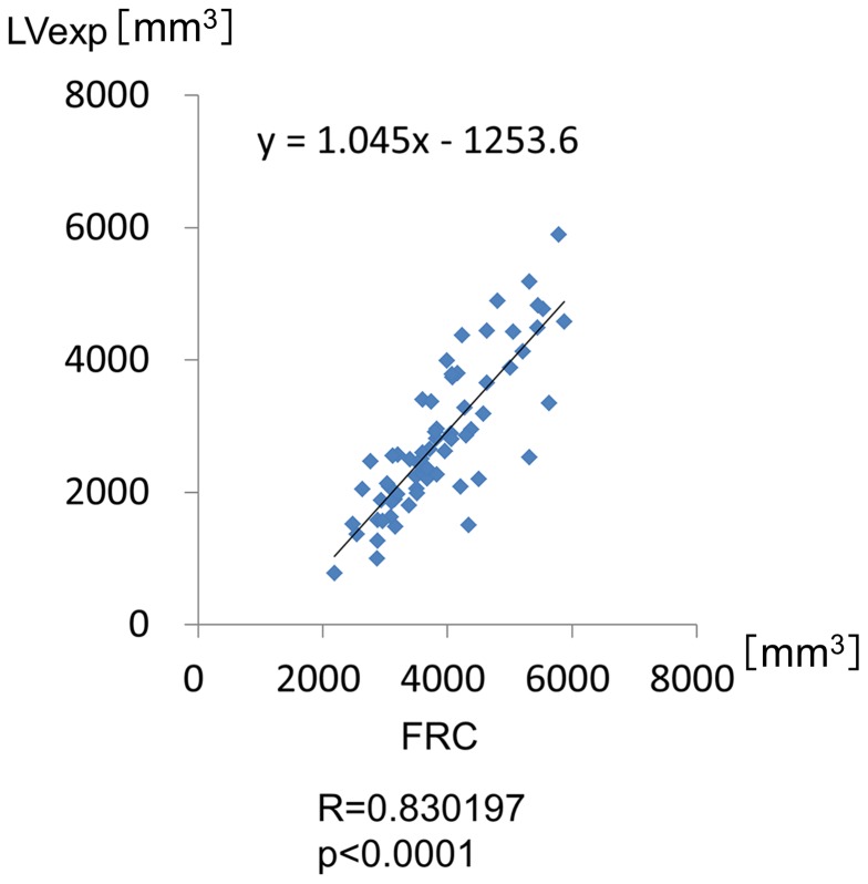
Sixty-seven subjects (15 at risk, 18, 20, and 14 for COPD stages 1, 2, and 3) underwent pulmonary function tests and computed tomography scans at full inspiration and expiration (at functional residual capacity). LV and eight selected identical airways were measured in the right lung. Ai was measured at the mid-portion of the 3(rd), the segmental bronchus, to 6(th) generation of the airways, leading to 32 measurements per subject.
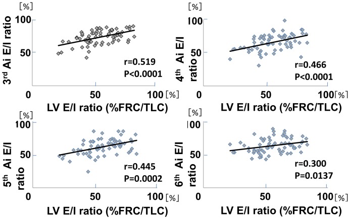
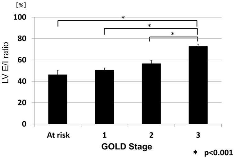
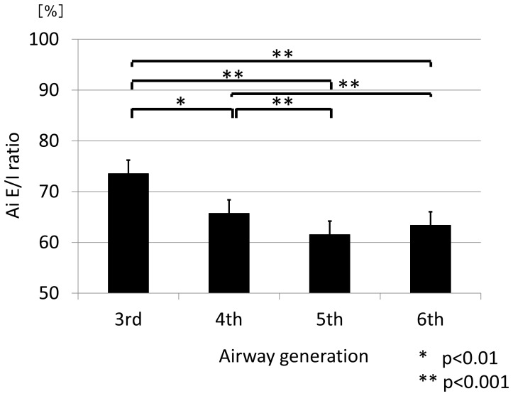
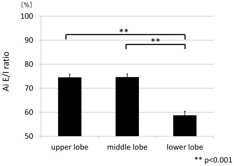
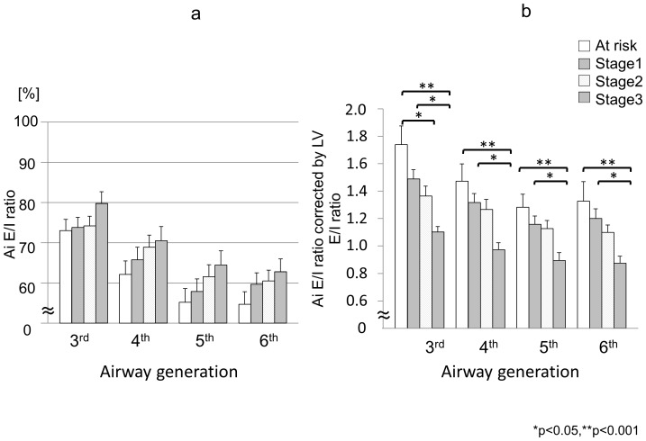
The ratio of expiratory to inspiratory LV (LV E/I ratio) and Ai (Ai E/I ratio) was defined for evaluation of changes. The LV E/I ratio increased as COPD severity progressed. As the LV E/I ratio was smaller, the Ai E/I ratio was smaller at any generation among the subjects. Overall, the Ai E/I ratios were significantly smaller at the 5(th) (61.5%) and 6(th) generations (63.4%) and than at the 3(rd) generation (73.6%, p<0.001 for each), and also significantly lower in the lower lobe than in the upper or middle lobe (p<0.001 for each). And, the Ai E/I ratio decreased as COPD severity progressed only when the ratio was corrected by the LV E/I ratio (at risk v.s. stage 3 p<0.001, stage 1 v.s. stage 3 p<0.05).
From full inspiration to expiration, the airway luminal area shrinks more at the distal airways compared with the proximal airways and in the lower lobe compared with the other lobes. Generally, the airways shrink more as COPD severity progresses, but this phenomenon becomes apparent only when lung volume change from inspiration to expiration is taken into account.
尽管气道管腔面积(Ai)受肺容积(LV)影响,但其具体影响方式尚不完全清楚。我们推测,LV对Ai的影响会因气道分级、肺叶以及慢性阻塞性肺疾病(COPD)严重程度的不同而有所差异。
67名受试者(15名有风险者,18名、20名和14名分别处于COPD 1、2和3期)在深吸气和呼气(功能残气量时)接受了肺功能测试和计算机断层扫描。测量了右肺的LV以及8条选定的相同气道。在气道的第3级(段支气管)至第6级的中部测量Ai,每位受试者共进行32次测量。
定义呼气与吸气LV之比(LV E/I比)和Ai之比(Ai E/I比)以评估变化情况。LV E/I比随着COPD严重程度的加重而增加。在所有受试者中,LV E/I比越小,任何分级的气道Ai E/I比就越小。总体而言,第5级(61.5%)和第6级(63.4%)气道的Ai E/I比显著小于第3级(73.6%,均p<0.001),下叶的Ai E/I比也显著低于上叶或中叶(均p<0.001)。并且,只有当Ai E/I比通过LV E/I比校正后,其才会随着COPD严重程度的加重而降低(有风险者与3期相比p<0.001,1期与3期相比p<0.05)。
从深吸气到呼气,与近端气道相比,远端气道的气道管腔面积缩小得更多,与其他肺叶相比,下叶的气道管腔面积缩小得更多。一般来说,随着COPD严重程度的加重,气道收缩得更多,但只有在考虑到从吸气到呼气的肺容积变化时,这种现象才会明显。