Zhai Chungang, Cheng Jing, Mujahid Haroon, Wang Hefeng, Kong Jing, Yin Yue, Li Jifu, Zhang Yun, Ji Xiaoping, Chen Wenqiang
The Key Laboratory of Cardiovascular Remodeling and Function Research, Chinese Ministry of Education and Chinese Ministry of Health, Shandong University Qilu Hospital, Jinan, Shandong, China.
PLoS One. 2014 Mar 5;9(3):e90563. doi: 10.1371/journal.pone.0090563. eCollection 2014.
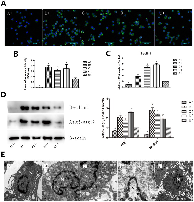
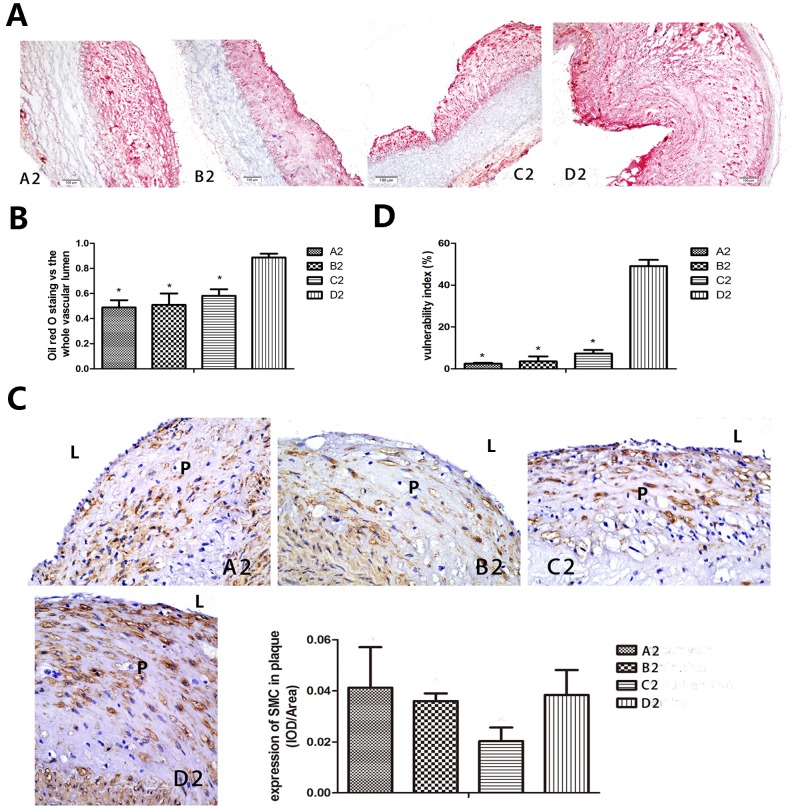
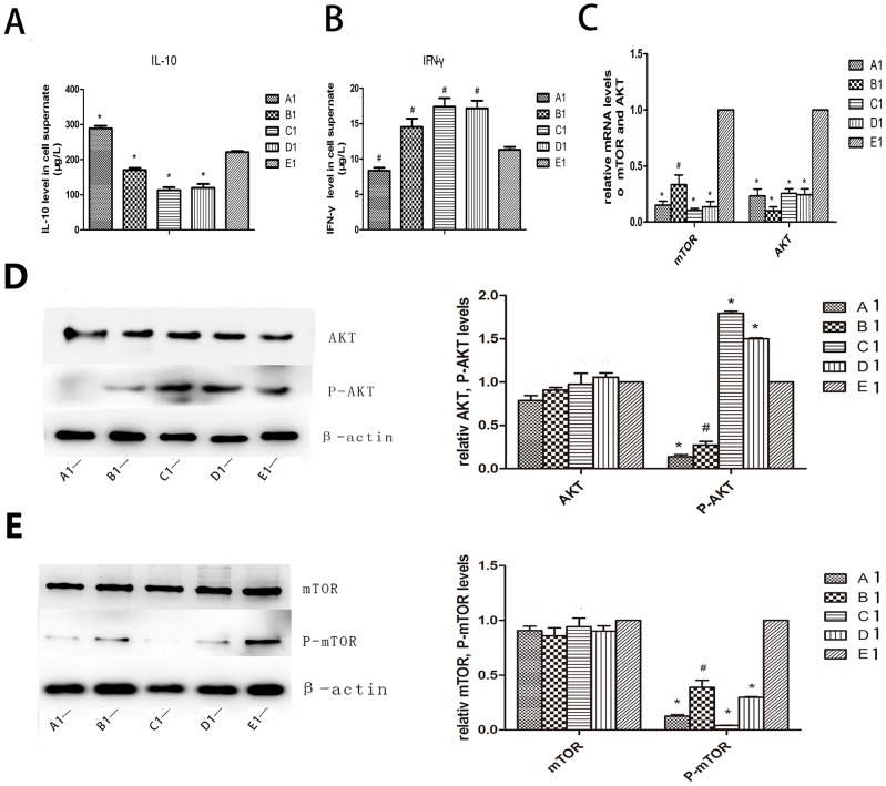
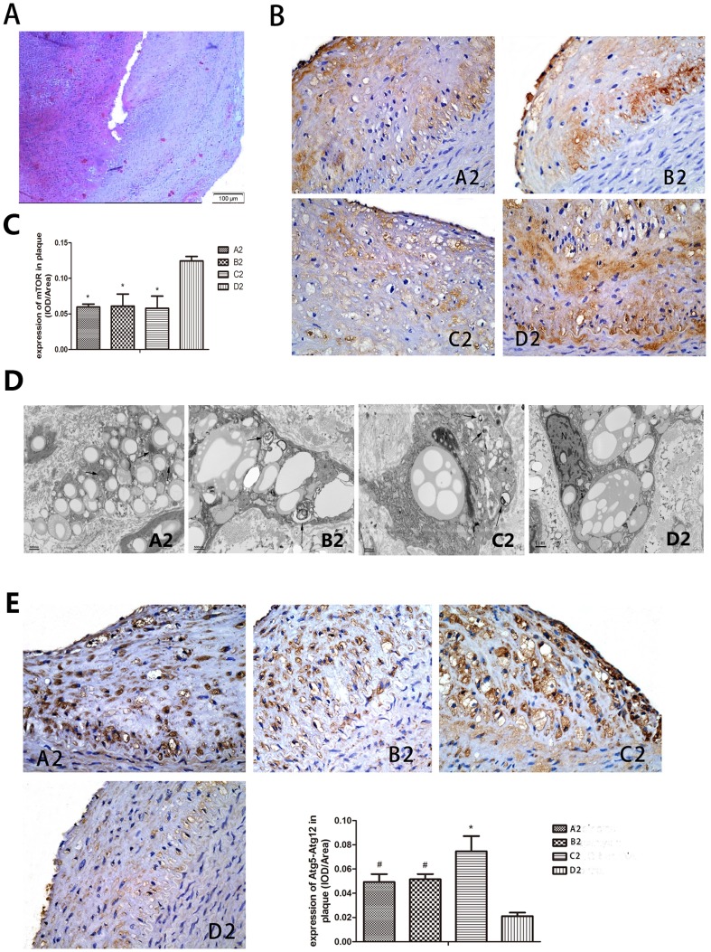
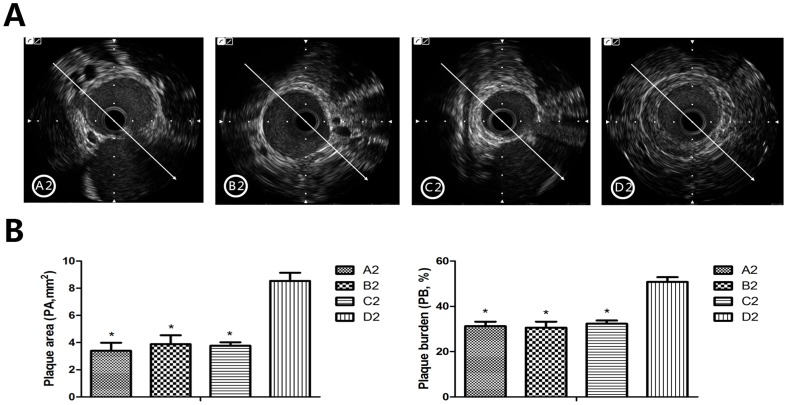
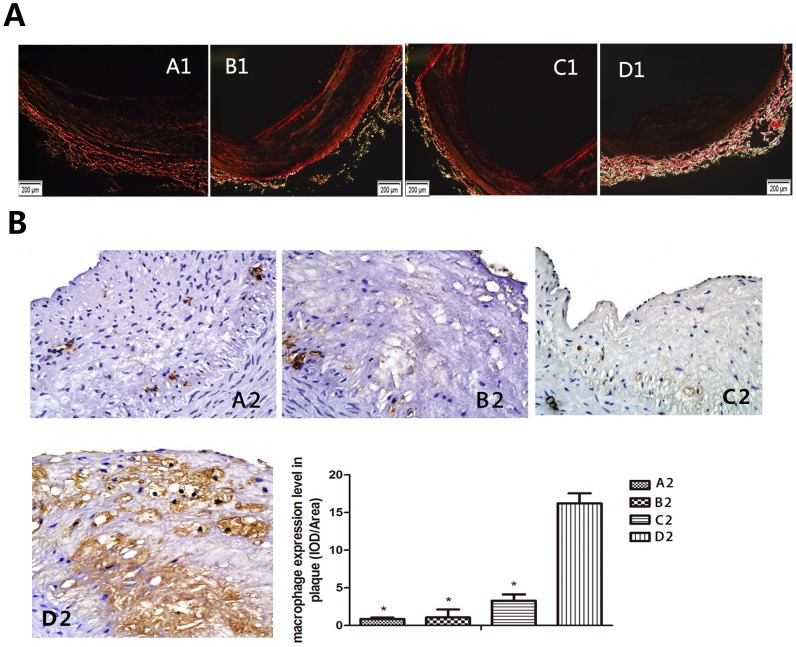
Macrophage infiltration contributes to the instability of atherosclerotic plaques. In the present study, we investigated whether selective inhibition of PI3K/Akt/mTOR signaling pathway can enhance the stability of atherosclerotic plaques by activation of macrophage autophagy. In vitro study, selective inhibitors or siRNA of PI3K/Akt/mTOR pathways were used to treat the rabbit's peritoneal primary macrophage cells. Inflammation related cytokines secreted by macrophages were measured. Ultrastructure changes of macrophages were examined by transmission electron microscope. mRNA or protein expression levels of autophagy related gene Beclin 1, protein 1 light chain 3 II dots (LC3-II) or Atg5-Atg12 conjugation were assayed by quantitative RT-PCR or Western blot. In vivo study, vulnerable plaque models were established in 40 New Zealand White rabbits and then drugs or siRNA were given for 8 weeks to inhibit the PI3K/Akt/mTOR signaling pathway. Intravascular ultrasound (IVUS) was performed to observe the plaque imaging. The ultrastructure of the abdominal aortic atherosclerosis lesions were analyzed with histopathology. RT-PCR or Western blot methods were used to measure the expression levels of corresponding autophagy related molecules. We found that macrophage autophagy was induced in the presence of Akt inhibitor, mTOR inhibitor and mTOR-siRNA in vitro study, while PI3K inhibitor had the opposite role. In vivo study, we found that macrophage autophagy increased significantly and the rabbits had lower plaque rupture incidence, lower plaque burden and decreased vulnerability index in the inhibitors or siRNA treated groups. We made a conclusion that selective inhibition of the Akt/mTOR signal pathway can reduce macrophages and stabilize the vulnerable atherosclerotic plaques by promoting macrophage autophagy.
巨噬细胞浸润会导致动脉粥样硬化斑块的不稳定。在本研究中,我们探究了选择性抑制PI3K/Akt/mTOR信号通路是否能够通过激活巨噬细胞自噬来增强动脉粥样硬化斑块的稳定性。在体外研究中,使用PI3K/Akt/mTOR通路的选择性抑制剂或小干扰RNA(siRNA)处理兔腹膜原代巨噬细胞。检测巨噬细胞分泌的炎症相关细胞因子。通过透射电子显微镜检查巨噬细胞的超微结构变化。采用定量逆转录聚合酶链反应(RT-PCR)或蛋白质免疫印迹法检测自噬相关基因Beclin 1、微管相关蛋白1轻链3Ⅱ型(LC3-II)的mRNA或蛋白表达水平,或Atg5-Atg12偶联物的表达水平。在体内研究中,在40只新西兰白兔中建立易损斑块模型,然后给予药物或siRNA 8周以抑制PI3K/Akt/mTOR信号通路。进行血管内超声(IVUS)观察斑块成像。用组织病理学分析腹主动脉粥样硬化病变的超微结构。采用RT-PCR或蛋白质免疫印迹法检测相应自噬相关分子的表达水平。我们发现在体外研究中,Akt抑制剂、mTOR抑制剂和mTOR-siRNA存在时可诱导巨噬细胞自噬,而PI3K抑制剂则起相反作用。在体内研究中,我们发现抑制剂或siRNA处理组巨噬细胞自噬显著增加,兔的斑块破裂发生率降低、斑块负荷降低且易损性指数降低。我们得出结论,选择性抑制Akt/mTOR信号通路可通过促进巨噬细胞自噬减少巨噬细胞并稳定易损动脉粥样硬化斑块。