Milks L C, Kumar N M, Houghten R, Unwin N, Gilula N B
Department of Molecular Biology, Research Institute of Scripps Clinic, La Jolla, CA 92037.
EMBO J. 1988 Oct;7(10):2967-75. doi: 10.1002/j.1460-2075.1988.tb03159.x.
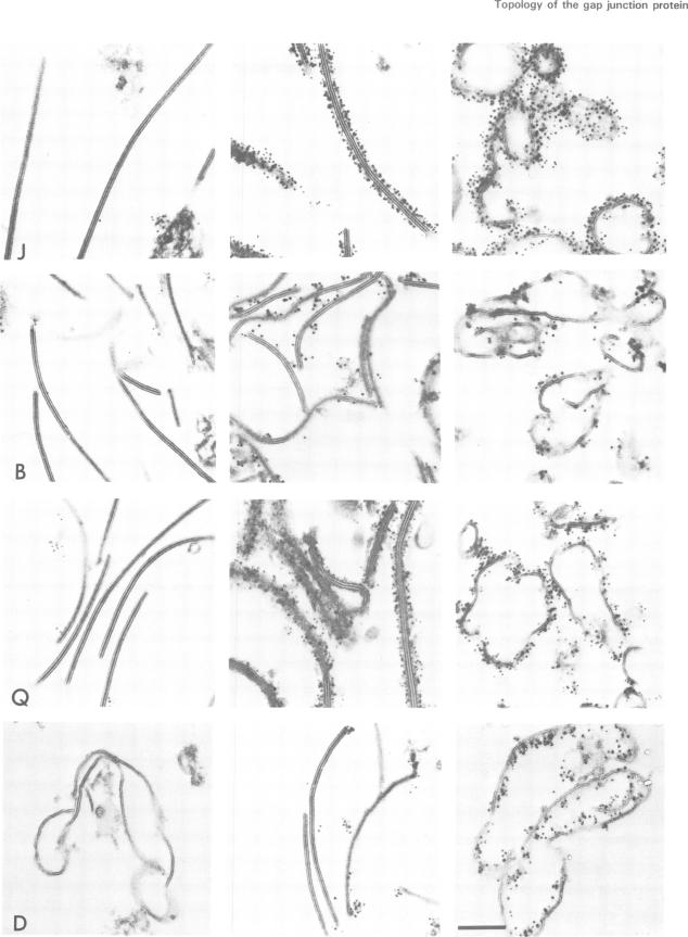
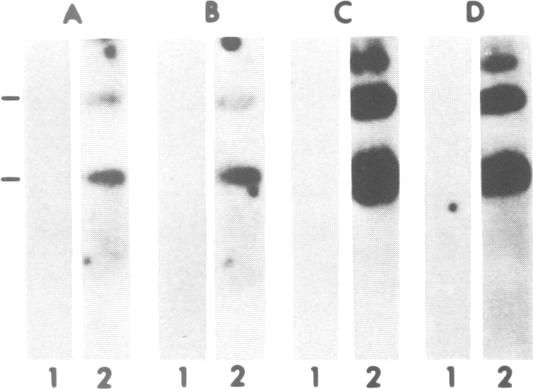
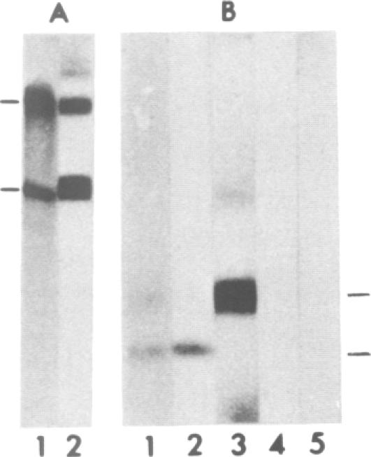
Synthetic peptides corresponding to sequences in the human liver gap junction protein were chemically synthesized and used for generation of peptide antisera to defined sequences in the protein. The antibodies were affinity purified and characterized by demonstrating that they specifically recognized both their corresponding synthetic peptide (as indicated by dot blot analysis) and the native 32-kd gap junction protein (by immunoblotting). The specificity of a subset of the different site-specific antibodies was subsequently confirmed by demonstration of their binding to specific gap junction fragments produced by treatment with a lysine-specific endoproteinase. Immunoelectron microscopy was used to localize the specific peptide antibody epitopes to either the cytoplasmic or extracellular surfaces of the gap junction. Results indicate a transmembrane orientation for the protein with the amino and carboxyl termini located on the cytoplasmic side of the membrane. Based on these data, a model is proposed for the transmembrane folding of the gap junction protein.
化学合成了与人类肝脏间隙连接蛋白序列相对应的合成肽,并用于生成针对该蛋白特定序列的肽抗血清。通过证明这些抗体能够特异性识别其相应的合成肽(如斑点印迹分析所示)和天然的32-kd间隙连接蛋白(通过免疫印迹),对抗体进行了亲和纯化和表征。随后,通过证明不同位点特异性抗体的一个子集与赖氨酸特异性内蛋白酶处理产生的特定间隙连接片段结合,证实了其特异性。免疫电子显微镜用于将特异性肽抗体表位定位到间隙连接的细胞质或细胞外表面。结果表明该蛋白具有跨膜取向,其氨基和羧基末端位于膜的细胞质侧。基于这些数据,提出了间隙连接蛋白跨膜折叠的模型。