• 文献检索
  • 文档翻译
  • 深度研究
  • 学术资讯
  • Suppr Zotero 插件Zotero 插件
  • 邀请有礼
  • 套餐&价格
  • 历史记录
应用&插件
Suppr Zotero 插件Zotero 插件浏览器插件Mac 客户端Windows 客户端微信小程序
定价
高级版会员购买积分包购买API积分包
服务
文献检索文档翻译深度研究API 文档MCP 服务
关于我们
关于 Suppr公司介绍联系我们用户协议隐私条款
关注我们

Suppr 超能文献

核心技术专利:CN118964589B侵权必究
粤ICP备2023148730 号-1Suppr @ 2026

文献检索

告别复杂PubMed语法,用中文像聊天一样搜索,搜遍4000万医学文献。AI智能推荐,让科研检索更轻松。

立即免费搜索

文件翻译

保留排版,准确专业,支持PDF/Word/PPT等文件格式,支持 12+语言互译。

免费翻译文档

深度研究

AI帮你快速写综述,25分钟生成高质量综述,智能提取关键信息,辅助科研写作。

立即免费体验

OLC1的过表达促进人食管鳞状细胞癌的肿瘤发生。

Overexpression of OLC1 promotes tumorigenesis of human esophageal squamous cell carcinoma.

作者信息

Li Xiao, Suo Jing, Shao Shujuan, Xue Liyan, Chen Wei, Dong Lijia, Shi Ji, Fu Ming, Lu Ning, Zhan Qimin, Tong Tong

机构信息

State Key Laboratory of Molecular Oncology, Cancer Institute & Cancer Hospital, Chinese Academy of Medical Sciences & Peking Union Medical College, Beijing, China; CAS Key Laboratory of Separation Science for Analytical Chemistry, Dalian Institute of Chemical Physics, Chinese Academy of Science, Dalian, Liaoning, China; Department of Histology and Embryology, Dalian Medical University, Dalian, Liaoning, China.

Department of Histology and Embryology, Dalian Medical University, Dalian, Liaoning, China.

出版信息

PLoS One. 2014 Mar 7;9(3):e90958. doi: 10.1371/journal.pone.0090958. eCollection 2014.

DOI:10.1371/journal.pone.0090958
PMID:24608342
原文链接:https://pmc.ncbi.nlm.nih.gov/articles/PMC3946619/
Abstract

PURPOSE

OLC1 was recently identified to be a potential oncogene. However, the role of OLC1 in human esophageal cell carcinoma (ESCC) is unknown. The aim of this study was therefore to evaluate the expression of OLC1 in human ESCC from normal, premalignant, and malignant lesions, and to clarify the mechanisms by which OLC1 contributes to the progression of ESCC.

EXPERIMENTAL DESIGN

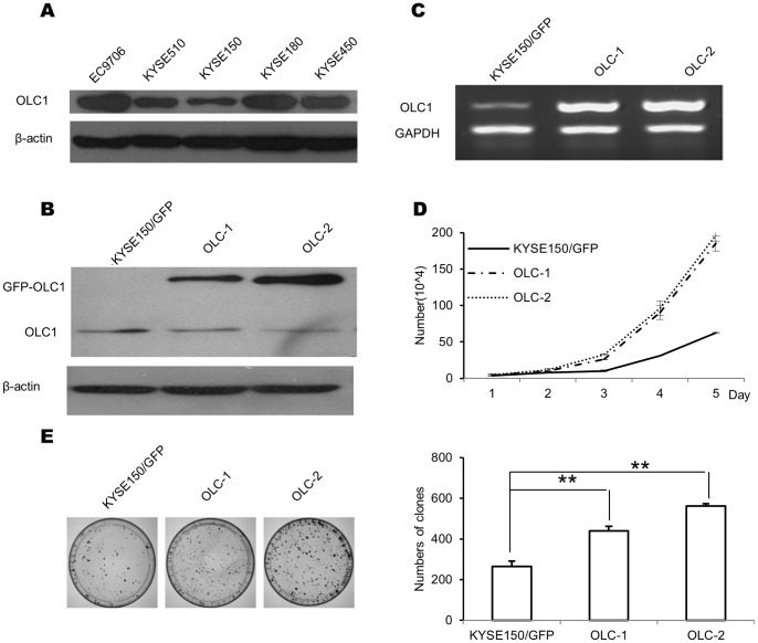
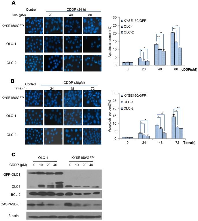
Two hundred and fourteen paired ESCC specimens, and an independent set from 28 ESCC patients, were used to analyze the correlation between OLC1 expression and the pathological characteristics of tumors using immunohistochemistry. Stable OLC1-overexpressing and OLC1-interfering esophageal cancer cells were established and a series of experimental methods were used to investigate the biological functions and mechanisms of action of OLC1.

RESULTS

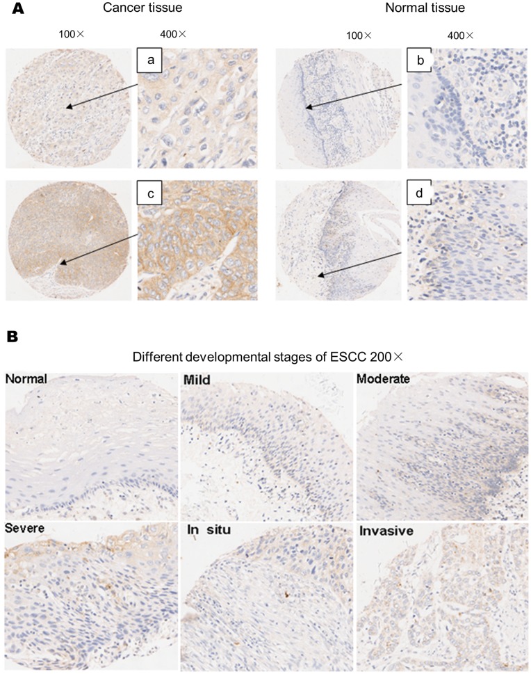
We showed that OLC1 was overexpressed in 145 of 214 (67.8%) of human ESCC specimens, compared with in only 59 of 214 (27.57%) paired adjacent normal tissues (P<0.001). OLC1 overexpression occurred at a rate of 35% (10/28) at the stage of mild/moderate dysplasia, but was significantly upregulated to 66% (22/33) at the stages of severe dysplasia and in situ carcinoma, while 71% positive staining (22/28) was observed in invasive carcinoma tissues compared with normal tissues (P<0.05). We also provided evidence that OLC1 abnormalities significantly altered the cell proliferation and apoptosis induced by cytotoxic agents. OLC1 overexpression suppressed apoptosis, and was associated with attenuated caspase-3 activation and increased Bcl-2 stability.

CONCLUSION

Our study provides strong evidence suggesting OLC1 abnormalities may contribute to the development of human ESCC and have some important clinical significance.

摘要

目的

OLC1最近被鉴定为一种潜在的癌基因。然而,OLC1在人类食管癌细胞癌(ESCC)中的作用尚不清楚。因此,本研究的目的是评估OLC1在人类ESCC正常、癌前和恶性病变中的表达,并阐明OLC1促进ESCC进展的机制。

实验设计

使用214对ESCC标本以及来自28例ESCC患者的独立样本,通过免疫组织化学分析OLC1表达与肿瘤病理特征之间的相关性。建立了稳定过表达OLC1和干扰OLC1的食管癌细胞,并采用一系列实验方法研究OLC1的生物学功能和作用机制。

结果

我们发现,在214例人类ESCC标本中,有145例(67.8%)OLC1过表达,而在214对配对的相邻正常组织中只有59例(27.57%)过表达(P<0.001)。在轻度/中度发育异常阶段,OLC1过表达率为35%(10/28),但在重度发育异常和原位癌阶段显著上调至66%(22/33),而在浸润癌组织中与正常组织相比观察到71%的阳性染色(22/28)(P<0.05)。我们还提供证据表明,OLC1异常显著改变了细胞毒性药物诱导的细胞增殖和凋亡。OLC1过表达抑制凋亡,并与caspase-3激活减弱和Bcl-2稳定性增加有关。

结论

我们的研究提供了有力证据,表明OLC1异常可能促进人类ESCC的发生发展,并具有重要的临床意义。

https://cdn.ncbi.nlm.nih.gov/pmc/blobs/62ee/3946619/03f4ae4f06ca/pone.0090958.g004.jpg
https://cdn.ncbi.nlm.nih.gov/pmc/blobs/62ee/3946619/1ded50128891/pone.0090958.g001.jpg
https://cdn.ncbi.nlm.nih.gov/pmc/blobs/62ee/3946619/af4dd9e0ae29/pone.0090958.g002.jpg
https://cdn.ncbi.nlm.nih.gov/pmc/blobs/62ee/3946619/27ed53632295/pone.0090958.g003.jpg
https://cdn.ncbi.nlm.nih.gov/pmc/blobs/62ee/3946619/03f4ae4f06ca/pone.0090958.g004.jpg

相似文献

1
Overexpression of OLC1 promotes tumorigenesis of human esophageal squamous cell carcinoma.OLC1的过表达促进人食管鳞状细胞癌的肿瘤发生。
PLoS One. 2014 Mar 7;9(3):e90958. doi: 10.1371/journal.pone.0090958. eCollection 2014.
2
L1CAM drives oncogenicity in esophageal squamous cell carcinoma by stimulation of ezrin transcription.L1CAM 通过刺激 ezrin 转录促进食管鳞癌的致癌性。
J Mol Med (Berl). 2017 Dec;95(12):1355-1368. doi: 10.1007/s00109-017-1595-4. Epub 2017 Sep 22.
3
[Overexpression of OLC1 in Lung Squamous Cell Carcinoma Tissues is Associated with Poor Prognosis of Patients].[OLC1在肺鳞状细胞癌组织中的过表达与患者预后不良相关]
Zhongguo Fei Ai Za Zhi. 2017 May 20;20(5):346-351. doi: 10.3779/j.issn.1009-3419.2017.05.08.
4
Protein and gene expression characteristics of heterogeneous nuclear ribonucleoprotein H1 in esophageal squamous cell carcinoma.食管鳞状细胞癌中异质性细胞核核糖核蛋白H1的蛋白质和基因表达特征
World J Gastroenterol. 2016 Aug 28;22(32):7322-31. doi: 10.3748/wjg.v22.i32.7322.
5
Long noncoding RNA, tissue differentiation-inducing nonprotein coding RNA is upregulated and promotes development of esophageal squamous cell carcinoma.长链非编码RNA,组织分化诱导非蛋白质编码RNA上调并促进食管鳞状细胞癌的发展。
Dis Esophagus. 2016 Nov;29(8):950-958. doi: 10.1111/dote.12436. Epub 2016 Feb 2.
6
miR‑378a‑3p exerts tumor suppressive function on the tumorigenesis of esophageal squamous cell carcinoma by targeting Rab10.miR-378a-3p 通过靶向 Rab10 对食管鳞状细胞癌的发生发挥肿瘤抑制作用。
Int J Mol Med. 2018 Jul;42(1):381-391. doi: 10.3892/ijmm.2018.3639. Epub 2018 Apr 24.
7
Overexpression of miR-191 Predicts Poor Prognosis and Promotes Proliferation and Invasion in Esophageal Squamous Cell Carcinoma.miR-191的过表达预示食管鳞状细胞癌的预后不良并促进其增殖和侵袭。
Yonsei Med J. 2017 Nov;58(6):1101-1110. doi: 10.3349/ymj.2017.58.6.1101.
8
Effect and mechanism of RUNX3 gene on biological characteristics of human esophageal squamous cell carcinoma (ESCC).RUNX3基因对人食管鳞状细胞癌(ESCC)生物学特性的影响及机制
Med Oncol. 2015 Jan;32(1):357. doi: 10.1007/s12032-014-0357-1. Epub 2014 Nov 27.
9
High expression of Collagen Triple Helix Repeat Containing 1 (CTHRC1) facilitates progression of oesophageal squamous cell carcinoma through MAPK/MEK/ERK/FRA-1 activation.含胶原蛋白三螺旋重复序列1(CTHRC1)的高表达通过激活丝裂原活化蛋白激酶/丝裂原活化蛋白激酶激酶/细胞外信号调节激酶/转录激活因子1(MAPK/MEK/ERK/FRA-1)促进食管鳞状细胞癌进展。
J Exp Clin Cancer Res. 2017 Jun 23;36(1):84. doi: 10.1186/s13046-017-0555-8.
10
Tissue microarray analysis reveals a tight correlation between protein expression pattern and progression of esophageal squamous cell carcinoma.组织微阵列分析显示,蛋白质表达模式与食管鳞状细胞癌的进展之间存在紧密关联。
BMC Cancer. 2006 Dec 22;6:296. doi: 10.1186/1471-2407-6-296.

引用本文的文献

1
Accessory ESCRT-III proteins are conserved and selective regulators of Rab11a-exosome formation.附属 ESCRT-III 蛋白是 Rab11a-外体形成的保守且有选择性的调节因子。
J Extracell Vesicles. 2023 Mar;12(3):e12311. doi: 10.1002/jev2.12311.
2
Regulation of OLC1 protein expression by the anaphase-promoting complex.后期促进复合物对OLC1蛋白表达的调控
Oncol Lett. 2019 Mar;17(3):2639-2646. doi: 10.3892/ol.2019.9881. Epub 2019 Jan 2.
3
[Overexpression of OLC1 in Lung Squamous Cell Carcinoma Tissues is Associated with Poor Prognosis of Patients].

本文引用的文献

1
MITD1 is recruited to midbodies by ESCRT-III and participates in cytokinesis.MITD1 通过 ESCRT-III 招募到中体并参与胞质分裂。
Mol Biol Cell. 2012 Nov;23(22):4347-61. doi: 10.1091/mbc.E12-04-0292. Epub 2012 Sep 26.
2
Spg20-/- mice reveal multimodal functions for Troyer syndrome protein spartin in lipid droplet maintenance, cytokinesis and BMP signaling.Spg20-/- 小鼠揭示了 Troyer 综合征蛋白 Spartin 在脂滴维持、胞质分裂和 BMP 信号传导中的多模式功能。
Hum Mol Genet. 2012 Aug 15;21(16):3604-18. doi: 10.1093/hmg/dds191. Epub 2012 May 22.
3
Oxymatrine induces human pancreatic cancer PANC-1 cells apoptosis via regulating expression of Bcl-2 and IAP families, and releasing of cytochrome c.
[OLC1在肺鳞状细胞癌组织中的过表达与患者预后不良相关]
Zhongguo Fei Ai Za Zhi. 2017 May 20;20(5):346-351. doi: 10.3779/j.issn.1009-3419.2017.05.08.
4
Nuclear overexpression of the overexpressed in lung cancer 1 predicts worse prognosis in gastric adenocarcinoma.肺癌中过表达的1在细胞核中过表达预示胃腺癌预后更差。
Oncotarget. 2017 Feb 7;8(6):9442-9450. doi: 10.18632/oncotarget.14217.
5
Interaction with CCNH/CDK7 facilitates CtBP2 promoting esophageal squamous cell carcinoma (ESCC) metastasis via upregulating epithelial-mesenchymal transition (EMT) progression.与CCNH/CDK7相互作用通过上调上皮-间质转化(EMT)进程促进CtBP2促进食管鳞状细胞癌(ESCC)转移。
Tumour Biol. 2015 Sep;36(9):6701-14. doi: 10.1007/s13277-015-3354-x. Epub 2015 Mar 29.
6
OLC1 is overexpressed in breast cancer and its expression correlates with poor patient survival.OLC1在乳腺癌中过表达,其表达与患者的不良生存相关。
Tumour Biol. 2014 Sep;35(9):8823-7. doi: 10.1007/s13277-014-2130-7. Epub 2014 Jun 1.
氧化苦参碱通过调节 Bcl-2 和 IAP 家族的表达以及细胞色素 c 的释放诱导人胰腺癌细胞 PANC-1 凋亡。
J Exp Clin Cancer Res. 2011 Jun 29;30(1):66. doi: 10.1186/1756-9966-30-66.
4
Valproic acid-induced DNA damage increases embryonic p27(KIP1) and caspase-3 expression: a mechanism for valproic-acid induced neural tube defects.丙戊酸诱导的 DNA 损伤增加胚胎 p27(KIP1)和 caspase-3 的表达:丙戊酸诱导神经管缺陷的一种机制。
Reprod Toxicol. 2011 Nov;32(3):255-60. doi: 10.1016/j.reprotox.2011.05.020. Epub 2011 Jun 17.
5
Resveratrol selectively induces DNA Damage, independent of Smad4 expression, in its efficacy against human head and neck squamous cell carcinoma.白藜芦醇对人头颈鳞癌细胞的疗效与其选择性诱导 DNA 损伤有关,而不依赖于 Smad4 表达。
Clin Cancer Res. 2011 Aug 15;17(16):5402-11. doi: 10.1158/1078-0432.CCR-11-1072. Epub 2011 Jun 24.
6
Cytotoxicity of tubeimoside I in human choriocarcinoma JEG-3 cells by induction of cytochrome c release and apoptosis via the mitochondrial-related signaling pathway.通过线粒体相关信号通路诱导细胞色素 c 释放和细胞凋亡,导致管状孢元苷 I 对人绒毛膜癌细胞 JEG-3 的细胞毒性。
Int J Mol Med. 2011 Oct;28(4):579-87. doi: 10.3892/ijmm.2011.727. Epub 2011 Jun 17.
7
Immunohistochemical evaluation of p53, FHIT, and IGF2 gene expression in esophageal cancer.食管癌中 p53、FHIT 和 IGF2 基因表达的免疫组化评估。
Dis Esophagus. 2012 Jan;25(1):81-7. doi: 10.1111/j.1442-2050.2011.01213.x. Epub 2011 Jun 10.
8
Gene mutations and polymorphisms of TP53 and FHIT in chronic esophagitis and esophageal carcinoma.TP53 和 FHIT 基因的突变和多态性与慢性食管炎和食管癌。
Anticancer Res. 2011 May;31(5):1685-90.
9
Using proteomic approach to identify tumor-associated proteins as biomarkers in human esophageal squamous cell carcinoma.采用蛋白质组学方法鉴定人食管鳞癌相关肿瘤标志物。
J Proteome Res. 2011 Jun 3;10(6):2863-72. doi: 10.1021/pr200141c. Epub 2011 May 3.
10
CyclinD1, p53, E-cadherin, and VEGF discordant expression in paired regional metastatic lymph nodes of esophageal squamous cell carcinoma: a tissue array analysis.食管鳞癌配对区域性转移淋巴结中细胞周期蛋白 D1、p53、E-钙黏蛋白和血管内皮生长因子的表达不一致:组织芯片分析。
J Surg Oncol. 2011 Sep 1;104(3):236-43. doi: 10.1002/jso.21921. Epub 2011 Apr 7.