Mandeville Joseph B, Liu Christina H, Vanduffel Wim, Marota John J A, Jenkins Bruce G
Athinoula A. Martinos Center for Biomedical Imaging, Massachusetts General Hospital, Charlestown, MA 02129, USA.
National Institute of Biomedical Imaging and Bioengineering, Bethesda, MD 20817, USA.
Neuropharmacology. 2014 Sep;84:65-78. doi: 10.1016/j.neuropharm.2014.02.018. Epub 2014 Mar 5.
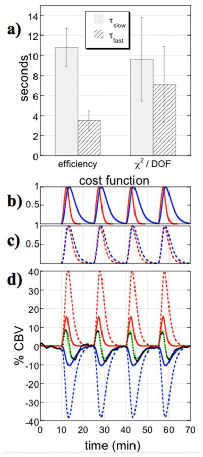
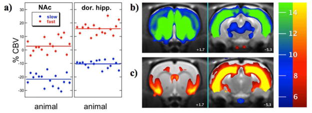
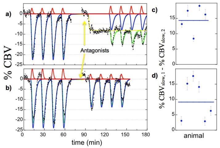
Although functional MRI traditionally has been applied mainly to study changes in task-induced brain function, evolving acquisition methodologies and improved knowledge of signal mechanisms have increased the utility of this method for studying responses to pharmacological stimuli, a technique often dubbed "phMRI". The proliferation of higher magnetic field strengths and the use of exogenous contrast agent have boosted detection power, a critical factor for successful phMRI due to the restricted ability to average multiple stimuli within subjects. Receptor-based models of neurovascular coupling, including explicit pharmacological models incorporating receptor densities and affinities and data-driven models that incorporate weak biophysical constraints, have demonstrated compelling descriptions of phMRI signal induced by dopaminergic stimuli. This report describes phMRI acquisition and analysis methodologies, with an emphasis on data-driven analyses. As an example application, statistically efficient data-driven regressors were used to describe the biphasic response to the mu-opioid agonist remifentanil, and antagonism using dopaminergic and GABAergic ligands revealed modulation of the mesolimbic pathway. Results illustrate the power of phMRI as well as our incomplete understanding of mechanisms underlying the signal. Future directions are discussed for phMRI acquisitions in human studies, for evolving analysis methodologies, and for interpretative studies using the new generation of simultaneous PET/MRI scanners. This article is part of the Special Issue Section entitled 'Neuroimaging in Neuropharmacology'.
尽管传统上功能磁共振成像主要应用于研究任务诱发的脑功能变化,但不断发展的采集方法和对信号机制的深入了解增加了该方法在研究药物刺激反应方面的实用性,这种技术常被称为“药物功能磁共振成像(phMRI)”。更高磁场强度的普及和外源性造影剂的使用提高了检测能力,由于在受试者体内对多个刺激进行平均的能力有限,这是phMRI成功的关键因素。基于受体的神经血管耦合模型,包括纳入受体密度和亲和力的明确药理模型以及纳入弱生物物理约束的数据驱动模型,已经对多巴胺能刺激诱发的phMRI信号进行了令人信服的描述。本报告描述了phMRI的采集和分析方法,重点是数据驱动分析。作为一个示例应用,使用统计高效的数据驱动回归器来描述对μ-阿片受体激动剂瑞芬太尼的双相反应,并且使用多巴胺能和γ-氨基丁酸能配体的拮抗作用揭示了中脑边缘通路的调节。结果说明了phMRI的强大功能以及我们对信号潜在机制的不完全理解。讨论了人类研究中phMRI采集、不断发展的分析方法以及使用新一代同步正电子发射断层扫描/磁共振成像(PET/MRI)扫描仪的解释性研究的未来方向。本文是名为“神经药理学中的神经成像”的特刊部分的一部分。