Bake Shameena, Selvamani Amutha, Cherry Jessica, Sohrabji Farida
Women's Health in Neuroscience Program, Department of Neuroscience and Experimental Therapeutics, Texas A&M University College of Medicine, Bryan, TX, United States of America.
PLoS One. 2014 Mar 11;9(3):e91427. doi: 10.1371/journal.pone.0091427. eCollection 2014.
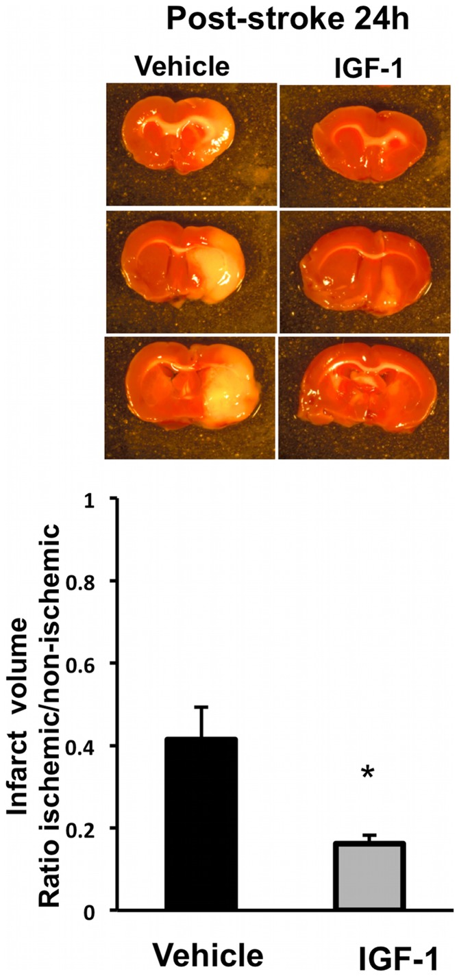
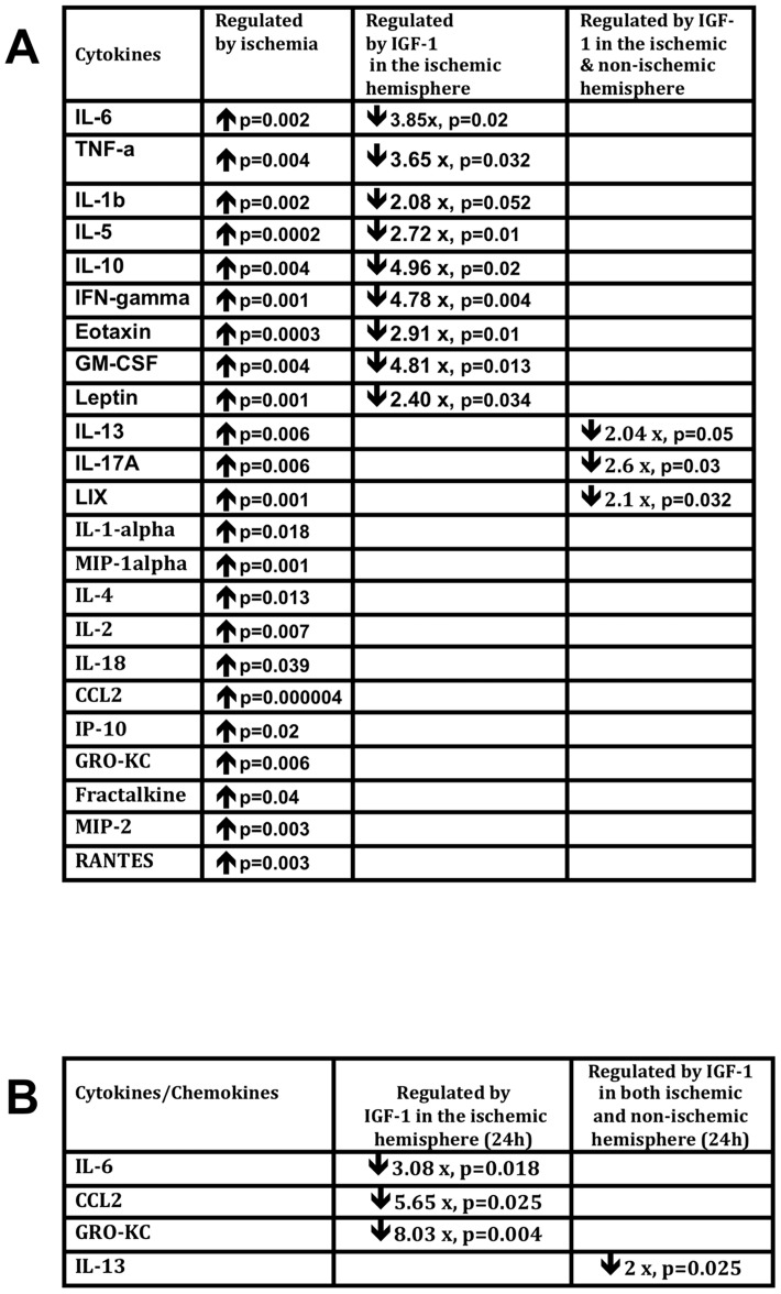
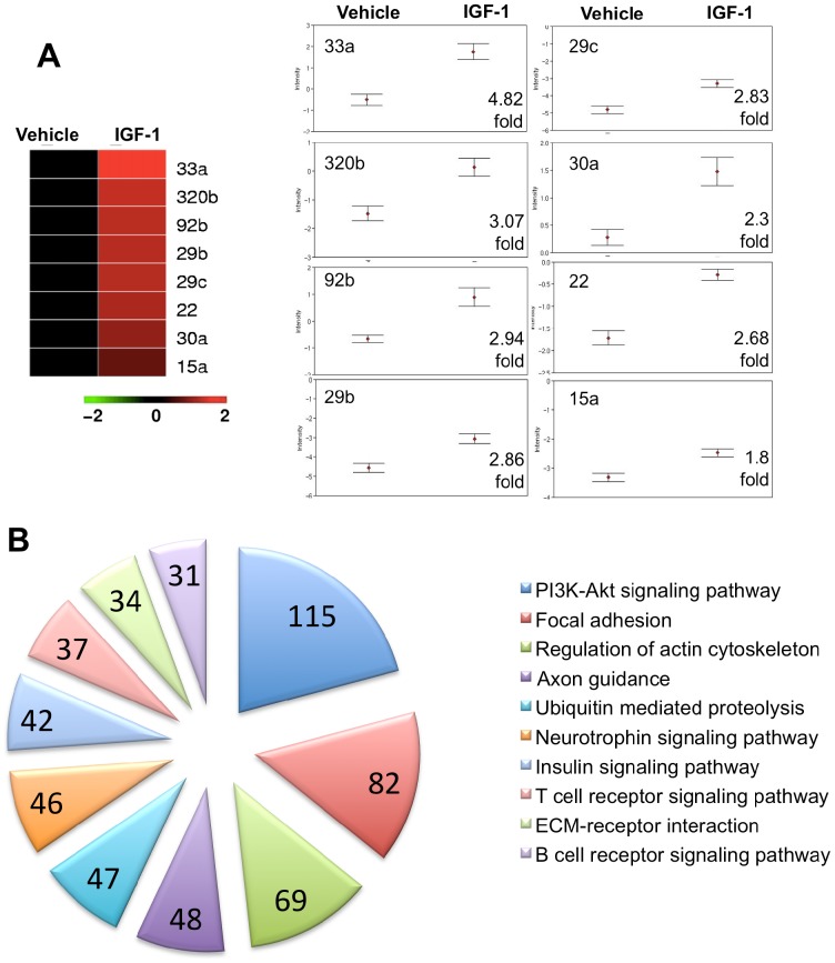
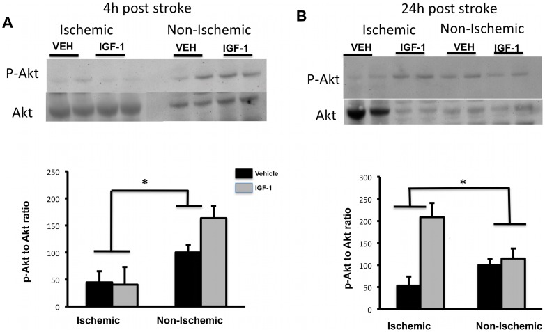
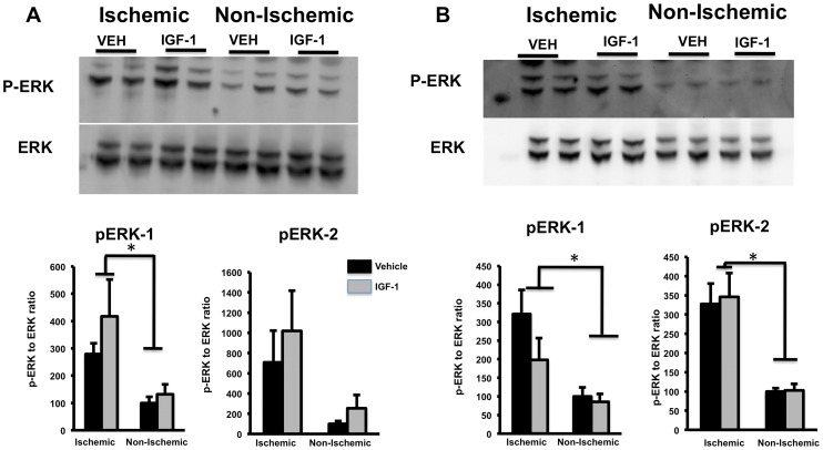
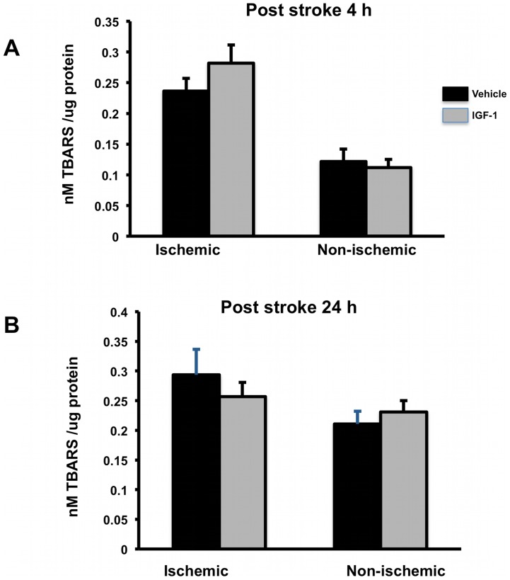
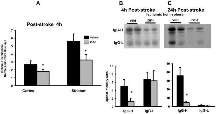
Ischemia-induced cerebral infarction is more severe in older animals as compared to younger animals, and is associated with reduced availability of insulin-like growth factor (IGF)-1. This study determined the effect of post-stroke IGF-1 treatment, and used microRNA profiling to identify mechanisms underlying IGF-1's neuroprotective actions. Post-stroke ICV administration of IGF-1 to middle-aged female rats reduced infarct volume by 39% when measured 24h later. MicroRNA analyses of ischemic tissue collected at the early post-stroke phase (4h) indicated that 8 out of 168 disease-related miRNA were significantly downregulated by IGF-1. KEGG pathway analysis implicated these miRNA in PI3K-Akt signaling, cell adhesion/ECM receptor pathways and T-and B-cell signaling. Specific components of these pathways were subsequently analyzed in vehicle and IGF-1 treated middle-aged females. Phospho-Akt was reduced by ischemia at 4h, but elevated by IGF-1 treatment at 24h. IGF-1 induced Akt activation was preceded by a reduction of blood brain barrier permeability at 4h post-stroke and global suppression of cytokines including IL-6, IL-10 and TNF-α. A subset of these cytokines including IL-6 was also suppressed by IGF-1 at 24h post-stroke. These data are the first to show that the temporal and mechanistic components of post-stroke IGF-1 treatment in older animals, and that cellular components of the blood brain barrier may serve as critical targets of IGF-1 in the aging brain.
与年轻动物相比,缺血诱导的脑梗死在老年动物中更为严重,并且与胰岛素样生长因子(IGF)-1的可用性降低有关。本研究确定了中风后IGF-1治疗的效果,并使用微小RNA谱分析来确定IGF-1神经保护作用的潜在机制。中风后对中年雌性大鼠进行脑室内注射IGF-1,24小时后测量发现梗死体积减少了39%。对中风后早期(4小时)收集的缺血组织进行微小RNA分析表明,168种与疾病相关的微小RNA中有8种被IGF-1显著下调。KEGG通路分析表明这些微小RNA参与了PI3K-Akt信号传导、细胞粘附/细胞外基质受体通路以及T细胞和B细胞信号传导。随后在给予载体和IGF-1治疗的中年雌性大鼠中分析了这些通路的特定成分。缺血在4小时时使磷酸化Akt减少,但IGF-1治疗在24小时时使其升高。IGF-1诱导的Akt激活之前,在中风后4小时血脑屏障通透性降低,并且包括IL-6、IL-10和TNF-α在内的细胞因子受到整体抑制。这些细胞因子中的一部分,包括IL-6,在中风后24小时也被IGF-1抑制。这些数据首次表明了老年动物中风后IGF-1治疗的时间和机制成分,并且血脑屏障的细胞成分可能是衰老大脑中IGF-1的关键靶点。