Olderøy Magnus Ø, Lilledahl Magnus B, Beckwith Marianne Sandvold, Melvik Jan Egil, Reinholt Finn, Sikorski Pawel, Brinchmann Jan E
The Norwegian Center for Stem Cell Research, Oslo University Hospital, Oslo, Norway.
Department of Physics, Norwegian University of Science and Technology, Trondheim, Norway.
PLoS One. 2014 Mar 13;9(3):e91662. doi: 10.1371/journal.pone.0091662. eCollection 2014.
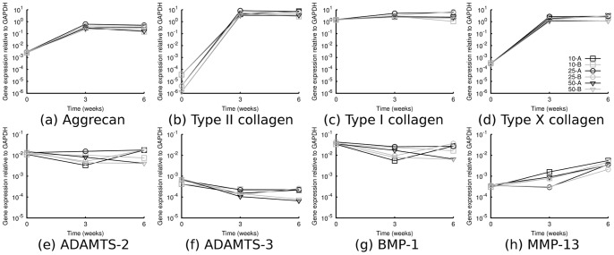
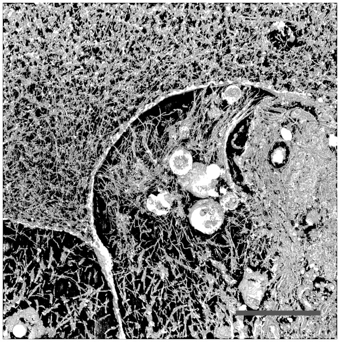
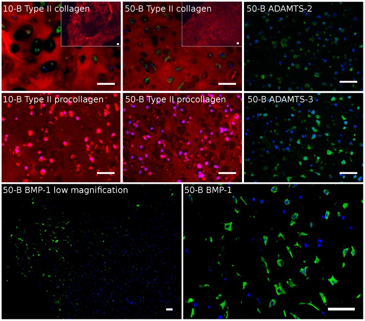
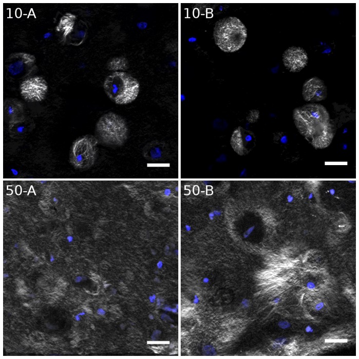
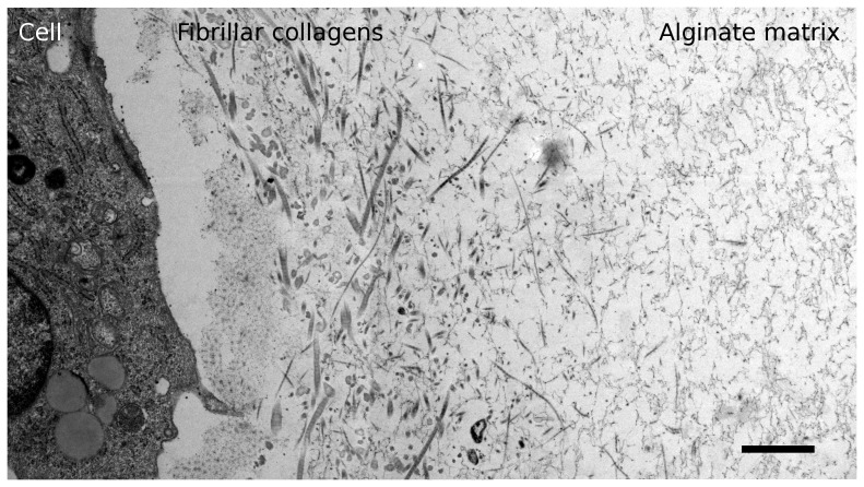
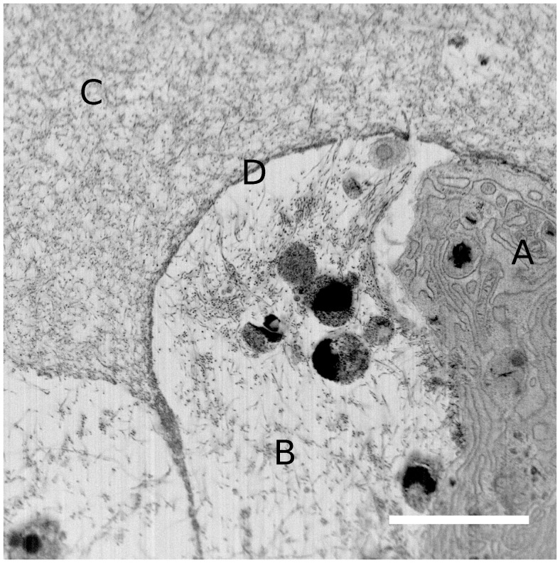
A popular approach to make neocartilage in vitro is to immobilize cells with chondrogenic potential in hydrogels. However, functional cartilage cannot be obtained by control of cells only, as function of cartilage is largely dictated by architecture of extracellular matrix (ECM). Therefore, characterization of the cells, coupled with structural and biochemical characterization of ECM, is essential in understanding neocartilage assembly to create functional implants in vitro. We focused on mesenchymal stem cells (MSC) immobilized in alginate hydrogels, and used immunohistochemistry (IHC) and gene expression analysis combined with advanced microscopy techniques to describe properties of cells and distribution and organization of the forming ECM. In particular, we used second harmonic generation (SHG) microscopy and focused ion beam/scanning electron microscopy (FIB/SEM) to study distribution and assembly of collagen. Samples with low cell seeding density (1e7 MSC/ml) showed type II collagen molecules distributed evenly through the hydrogel. However, SHG microscopy clearly indicated only pericellular localization of assembled fibrils. Their distribution was improved in hydrogels seeded with 5e7 MSC/ml. In those samples, FIB/SEM with nm resolution was used to visualize distribution of collagen fibrils in a three dimensional network extending from the pericellular region into the ECM. In addition, distribution of enzymes involved in procollagen processing were investigated in the alginate hydrogel by IHC. It was discovered that, at high cell seeding density, procollagen processing and fibril assembly was also occurring far away from the cell surface, indicating sufficient transport of procollagen and enzymes in the intercellular space. At lower cell seeding density, the concentration of enzymes involved in procollagen processing was presumably too low. FIB/SEM and SHG microscopy combined with IHC localization of specific proteins were shown to provide meaningful insight into ECM assembly of neocartilage, which will lead to better understanding of cartilage formation and development of new tissue engineering strategies.
一种在体外制造新软骨的常用方法是将具有软骨形成潜力的细胞固定在水凝胶中。然而,仅通过控制细胞无法获得功能性软骨,因为软骨的功能很大程度上取决于细胞外基质(ECM)的结构。因此,对细胞进行表征,同时结合ECM的结构和生化表征,对于理解新软骨组装以在体外创建功能性植入物至关重要。我们专注于固定在藻酸盐水凝胶中的间充质干细胞(MSC),并使用免疫组织化学(IHC)和基因表达分析,结合先进的显微镜技术来描述细胞的特性以及形成的ECM的分布和组织。特别是,我们使用二次谐波产生(SHG)显微镜和聚焦离子束/扫描电子显微镜(FIB/SEM)来研究胶原蛋白的分布和组装。低细胞接种密度(1×10⁷ MSC/ml)的样品显示II型胶原蛋白分子均匀分布在水凝胶中。然而,SHG显微镜清楚地表明组装的纤维仅位于细胞周围。在接种5×10⁷ MSC/ml的水凝胶中,它们的分布得到了改善。在这些样品中,使用具有纳米分辨率的FIB/SEM来可视化胶原蛋白纤维在从细胞周围区域延伸到ECM的三维网络中的分布。此外,通过IHC研究了藻酸盐水凝胶中参与前胶原加工的酶的分布。结果发现,在高细胞接种密度下,前胶原加工和纤维组装也发生在远离细胞表面的地方,这表明前胶原和酶在细胞间空间中有足够的运输。在较低的细胞接种密度下,参与前胶原加工的酶的浓度可能过低。结果表明,FIB/SEM和SHG显微镜与特定蛋白质的IHC定位相结合,能够为新软骨的ECM组装提供有意义的见解,这将有助于更好地理解软骨形成并开发新的组织工程策略。