Department of Neurology, The 105th Hospital of PLA, Clinic College, Anhui Medical University, Hefei 230031, China.
BMC Neurosci. 2014 Mar 17;15:41. doi: 10.1186/1471-2202-15-41.
Because there is little research on the effects of transplanted stem cells on neuronal metabolites in infarct areas, we transplanted human umbilical cord blood mesenchymal stem cells (hUCB-MSCs) into cerebral ischemic rabbits and examined the neuronal metabolites.
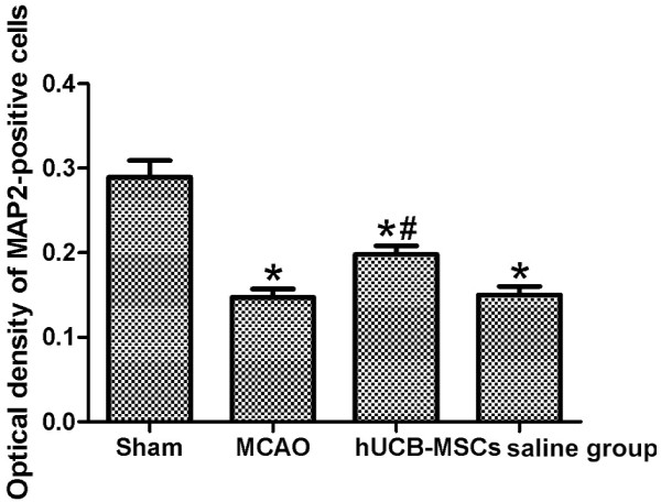
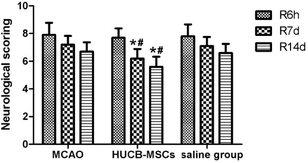
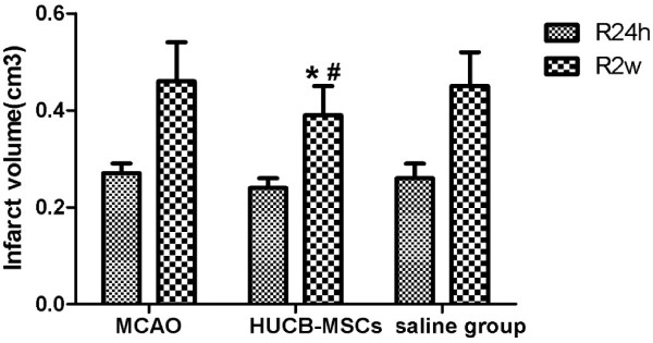
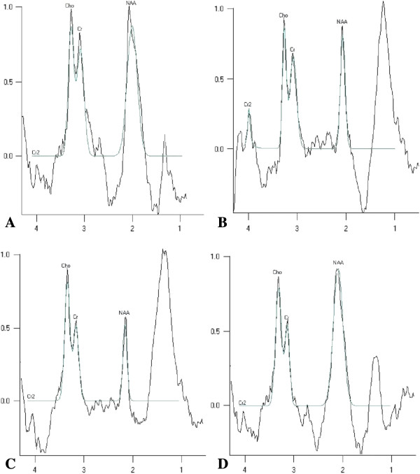
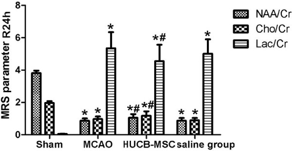
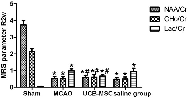
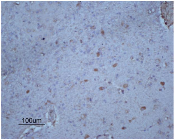
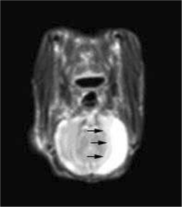
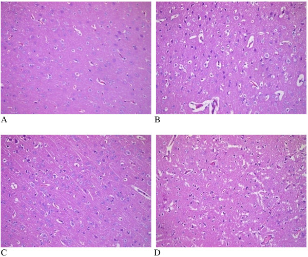
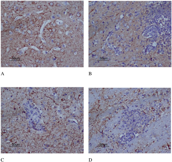
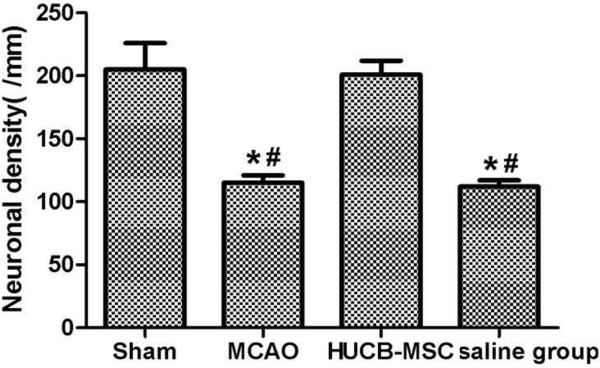
Rabbits (n = 40) were equally divided into sham, middle cerebral artery occlusion (MCAO), hUCB-MSC, and saline groups. The rabbit ischemic model was established by MCAO. The effects of hUCB-MSC transplantation were assessed by proton magnetic resonance spectroscopy (1H-MRS), neurological severity scores (NSSs), infarct area volume, neuronal density, and optical density (OD) of microtubule-associated protein 2 (MAP2)-positive cells. We also evaluated complete blood cell counts(CBCs) and serum biochemical parameters. NSSs in the hUCB-MSC group at 7 and 14 days after reperfusion were lower than in MCAO and saline groups (p < 0.05). Compared with MCAO and saline groups at 2 weeks after MCAO, the infarction volume in the hUCB-MSC group had decreased remarkably (p < 0.05). Significant neuronal metabolic changes occurred in the infarct area at 24 h and 2 weeks after MCAO. 1H-MRS revealed an elevation in the lactate (Lac)/creatine including phosphocreatine (Cr) ratio and a decrease in the N-acetylaspartate (NAA)/Cr and choline-containing phospholipids (Cho)/Cr ratios at 24 h after MCAO in the MCAO group (p < 0.01). Compared with saline and MCAO groups at 24 h and 2 weeks after MCAO, NAA/Cr and Cho/Cr ratios had increased significantly, whereas the Lac/Cr ratio had decreased significantly in the hUCB-MSC group (p < 0.01). Neuronal density and OD of MAP2-positive cells in the MCAO group were significantly lower than those in the sham group, whereas the neuronal density and OD of MAP2-positive cells in the hUCB-MSC group were higher than those in MCAO and saline groups (p < 0.05). CBCs and biochemical parameters were unchanged in the MCAO group at 24 h and 2 weeks after hUCB-MSC transplantation.
Transplanted hUCB-MSCs might ameliorate ischemic damage by influencing neuronal metabolites in the infarct area, providing additional evidence for neuroprotection by stem cells. No significant changes were observed in CBCs or serum biochemical parameters, suggesting that intravenous infusion of hUCB-MSCs is safe for rabbits in the short-term.
由于关于移植的干细胞对梗死区域神经元代谢物影响的研究较少,我们将人脐带血间充质干细胞(hUCB-MSCs)移植到脑缺血兔体内,并检测神经元代谢物。
将 40 只兔子等分为假手术组、大脑中动脉闭塞(MCAO)组、hUCB-MSC 组和盐水组。通过 MCAO 建立兔缺血模型。通过质子磁共振波谱(1H-MRS)、神经严重程度评分(NSSs)、梗死面积体积、神经元密度和微管相关蛋白 2(MAP2)阳性细胞的光密度(OD)评估 hUCB-MSC 移植的效果。还评估了全血细胞计数(CBC)和血清生化参数。再灌注后 7 天和 14 天,hUCB-MSC 组的 NSS 评分低于 MCAO 组和盐水组(p<0.05)。与 MCAO 组和盐水组在 MCAO 后 2 周相比,hUCB-MSC 组的梗死体积明显减小(p<0.05)。MCAO 后 24 小时和 2 周,梗死区发生明显的神经元代谢变化。1H-MRS 显示 MCAO 后 24 小时,MCAO 组乳酸(Lac)/肌酸(Cr)比值升高,N-乙酰天冬氨酸(NAA)/Cr 和胆碱含磷磷脂(Cho)/Cr 比值降低(p<0.01)。与 MCAO 组和盐水组在 MCAO 后 24 小时和 2 周相比,hUCB-MSC 组 NAA/Cr 和 Cho/Cr 比值显著升高,Lac/Cr 比值显著降低(p<0.01)。MCAO 组神经元密度和 MAP2 阳性细胞 OD 明显低于假手术组,hUCB-MSC 组神经元密度和 MAP2 阳性细胞 OD 明显高于 MCAO 组和盐水组(p<0.05)。hUCB-MSC 移植后 24 小时和 2 周,MCAO 组的 CBC 和生化参数无明显变化。
移植的 hUCB-MSCs 可能通过影响梗死区的神经元代谢物来改善缺血损伤,为干细胞的神经保护作用提供了更多证据。CBC 和血清生化参数无明显变化,提示短期内静脉输注 hUCB-MSCs 对兔是安全的。