Department of Pathobiology, Faculty of Veterinary Medicine, Tehran University, Tehran, Iran.
Diagn Pathol. 2014 Mar 17;9:59. doi: 10.1186/1746-1596-9-59.
Today, finding an ideal biomaterial to treat the large bone defects, delayed unions and non-unions remains a challenge for orthopaedic surgeions and researchers. Several studies have been carried out on the subject of bone regeneration, each having its own advantages. The present study has been designed in vivo to evaluate the effects of cellular auto-transplantation of tail vertebrae on healing of experimental critical bone defect in a dog model.
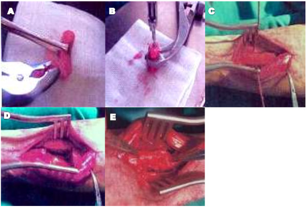
Six indigenous breeds of dog with 32 ± 3.6 kg average weight from both sexes (5 males and 1 female) received bilateral critical-sized ulnar segmental defects. After determining the health condition, divided to 2 groups: The Group I were kept as control I (n = 1) while in Group II (experimental group; n = 5) bioactive bone implants were inserted. The defects were implanted with either autogeneic coccygeal bone grafts in dogs with 3-4 cm diaphyseal defects in the ulna. Defects were stabilized with internal plate fixation, and the control defects were not stabilized. Animals were euthanized at 16 weeks and analyzed by histopathology.
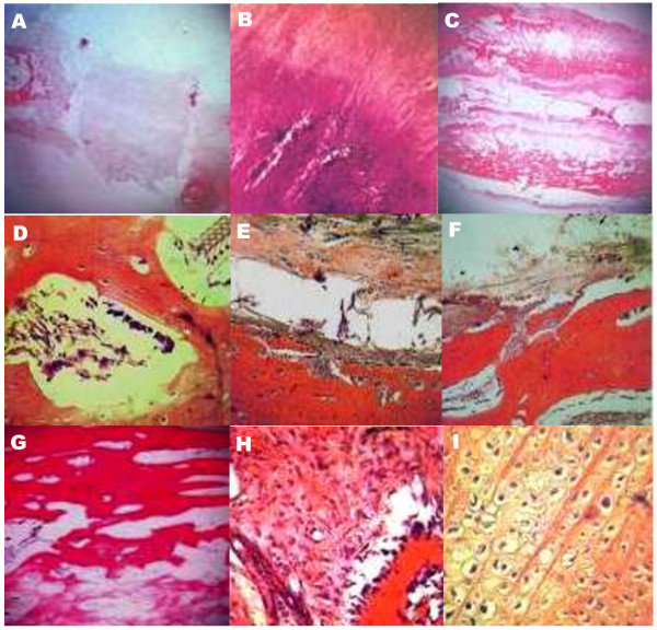
Histological evaluation of this new bone at sixteen weeks postoperatively revealed primarily lamellar bone, with the formation of new cortices and normal-appearing marrow elements. And also reformation cortical compartment and reconstitution of marrow space were observed at the graft-host interface together with graft resorption and necrosis responses. Finally, our data were consistent with the osteoconducting function of the tail autograft.
Our results suggested that the tail vertebrae autograft seemed to be a new source of autogenous cortical bone in order to supporting segmental long bone defects in dogs. Furthermore, cellular autotransplantation was found to be a successful replacement for the tail vertebrae allograft bone at 3-4 cm segmental defects in the canine mid- ulna. Clinical application using graft expanders or bone autotransplantation should be used carefully and requires further investigation.
The virtual slide(s) for this article can be found here: http://www.diagnosticpathology.diagnomx.eu/vs/2028232688119271.
如今,寻找理想的生物材料来治疗大的骨缺损、延迟愈合和不愈合仍然是骨科医生和研究人员面临的挑战。已经有多项关于骨再生的研究,每一项都有其自身的优势。本研究旨在体内评估尾椎细胞自体移植对犬模型实验性临界骨缺损愈合的影响。
选择 6 只平均体重为 32±3.6kg 的本地品种犬(5 雄 1 雌),进行双侧尺骨干节段性临界缺损。在确定健康状况后,将其分为 2 组:第 I 组为对照组 I(n=1),第 II 组(实验组;n=5)植入生物活性骨植入物。在 3-4cm 骨干缺损的犬中,将尾骨自体骨移植到尺骨缺损处。用内置钢板固定,对照组不固定。16 周后处死动物,进行组织病理学分析。
术后 16 周的新骨组织学评价显示主要为板层骨,形成新的皮质,并出现正常的骨髓成分。还观察到移植物-宿主界面的皮质腔再形成和骨髓腔重建,同时伴有移植物吸收和坏死反应。最终,我们的数据与尾骨自体移植物的成骨作用一致。
我们的结果表明,尾椎自体移植物似乎是一种新的自体皮质骨来源,可用于支撑犬的长骨骨干缺损。此外,在犬尺骨干 3-4cm 节段性缺损中,细胞自体移植被发现是尾骨同种异体骨移植的成功替代物。使用移植物扩张器或骨自体移植的临床应用应谨慎,并需要进一步研究。
本文的虚拟幻灯片可以在此处找到:http://www.diagnosticpathology.diagnomx.eu/vs/2028232688119271。