Bhattacharya Soumya, Dhar Purbarun, Das Sarit K, Ganguly Ranjan, Webster Thomas J, Nayar Suprabha
Biomaterials Group, Materials Science and Technology Division, CSIR-National Metallurgical Laboratory, Jamshedpur.
Nanofluids, Microfluidics and Bio-MEMS Laboratory, Department of Mechanical Engineering, Indian Institute of Technology-Madras, Chennai.
Int J Nanomedicine. 2014 Mar 10;9:1287-98. doi: 10.2147/IJN.S57122. eCollection 2014.
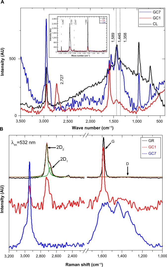
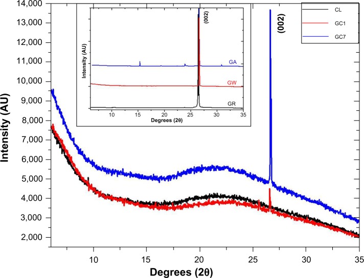
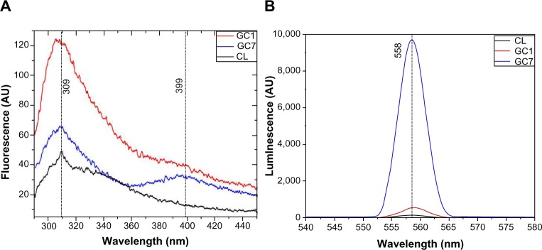
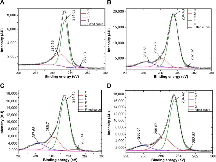
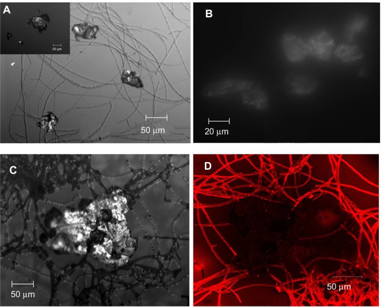
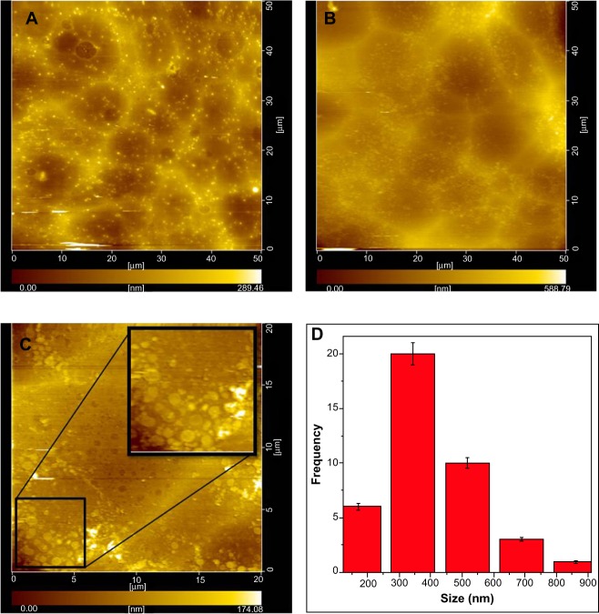
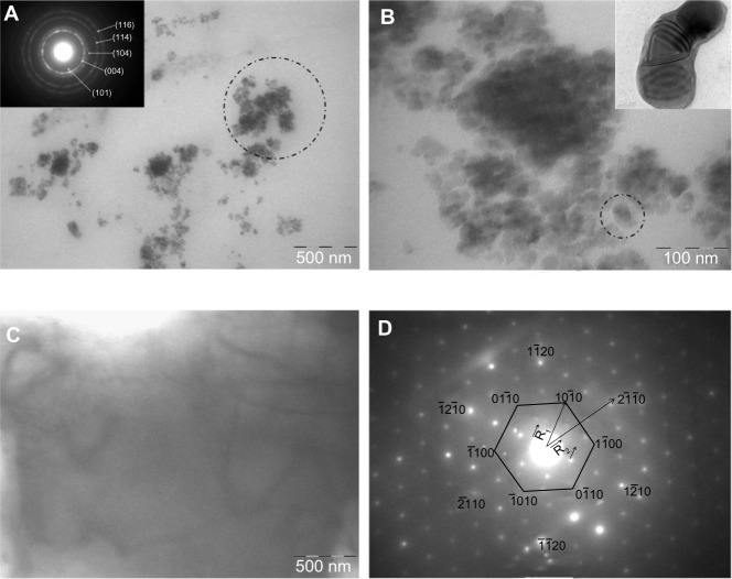
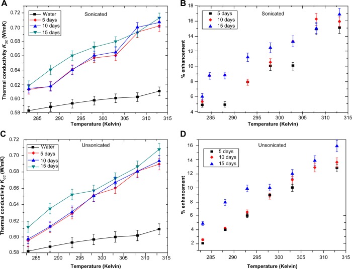
In the present study, the exfoliation of natural graphite (GR) directly to colloidal GR/graphene (G) nanostructures using collagen (CL) was studied as a safe and scalable process, akin to numerous natural processes and hence can be termed "biomimetic". Although the exfoliation and functionalization takes place in just 1 day, it takes about 7 days for the nano GR/G flakes to stabilize. The predominantly aromatic residues of the triple helical CL forms its own special micro and nanoarchitecture in acetic acid dispersions. This, with the help of hydrophobic and electrostatic forces, interacts with GR and breaks it down to nanostructures, forming a stable colloidal dispersion. Surface enhanced Raman spectroscopy, X-ray diffraction, photoluminescence, fluorescence, and X-ray photoelectron spectroscopy of the colloid show the interaction between GR and CL on day 1 and 7. Differential interference contrast images in the liquid state clearly reveal how the GR flakes are entrapped in the CL fibrils, with a corresponding fluorescence image showing the intercalation of CL within GR. Atomic force microscopy of graphene-collagen coated on glass substrates shows an average flake size of 350 nm, and the hexagonal diffraction pattern and thickness contours of the G flakes from transmission electron microscopy confirm ≤ five layers of G. Thermal conductivity of the colloid shows an approximate 17% enhancement for a volume fraction of less than approximately 0.00005 of G. Thus, through the use of CL, this new material and process may improve the use of G in terms of biocompatibility for numerous medical applications that currently employ G, such as internally controlled drug-delivery assisted thermal ablation of carcinoma cells.
在本研究中,研究了使用胶原蛋白(CL)将天然石墨(GR)直接剥离为胶体GR/石墨烯(G)纳米结构的过程,该过程安全且可扩展,类似于许多自然过程,因此可称为“仿生”。尽管剥离和功能化仅需1天时间,但纳米GR/G薄片大约需要7天才能稳定下来。三螺旋CL的主要芳香族残基在醋酸分散体中形成了其自身特殊的微纳米结构。借助疏水和静电力,它与GR相互作用并将其分解为纳米结构,形成稳定的胶体分散体。胶体的表面增强拉曼光谱、X射线衍射、光致发光、荧光和X射线光电子能谱显示了第1天和第7天GR与CL之间的相互作用。液态下的微分干涉对比图像清楚地揭示了GR薄片如何被困在CL原纤维中,相应的荧光图像显示了CL在GR中的嵌入。涂覆在玻璃基板上的石墨烯-胶原蛋白的原子力显微镜显示平均薄片尺寸为350nm,透射电子显微镜下G薄片的六边形衍射图案和厚度轮廓证实G≤五层。对于体积分数小于约0.00005的G,胶体的热导率显示出约17%的提高。因此,通过使用CL,这种新材料和工艺可能会改善G在众多目前使用G的医疗应用中的生物相容性,例如癌细胞的内部控制药物递送辅助热消融。