Suppr超能文献

磁共振成像在手臂海绵状血管瘤诊断与评估中的作用

Role of MRI in the diagnosis and evaluation of cavernous hemangioma of the arm.

作者信息

Ciurea M E, Bondari S, Stoica L E, Gheonea I A

机构信息

Department of Plastic and Reconstructive Surgery, Craiova University of Medicine and Pharmacy.

Radiology and Imaging Department, Craiova University of Medicine and Pharmacy.

出版信息

J Med Life. 2014 Mar 15;7(1):46-50. Epub 2014 Mar 25.

Abstract

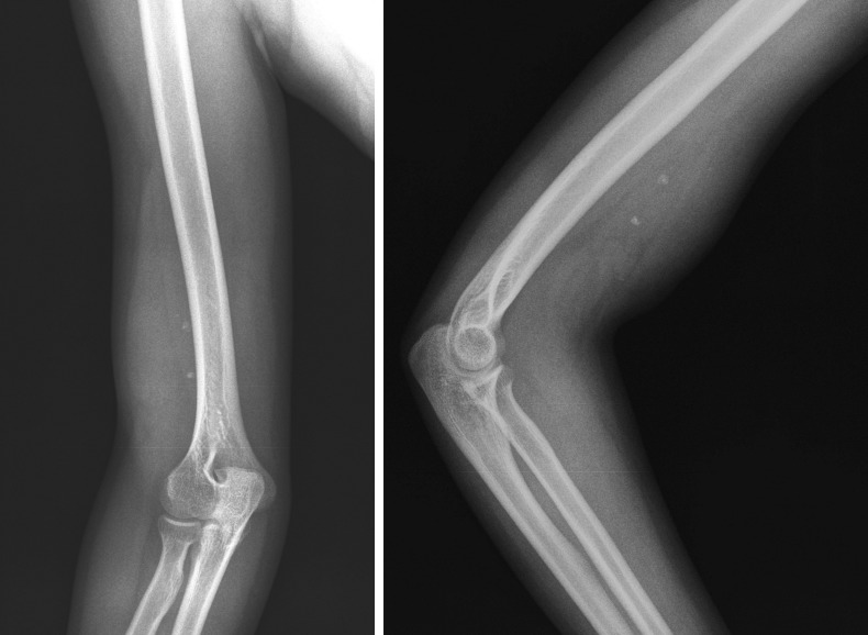
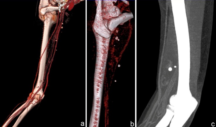
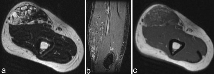
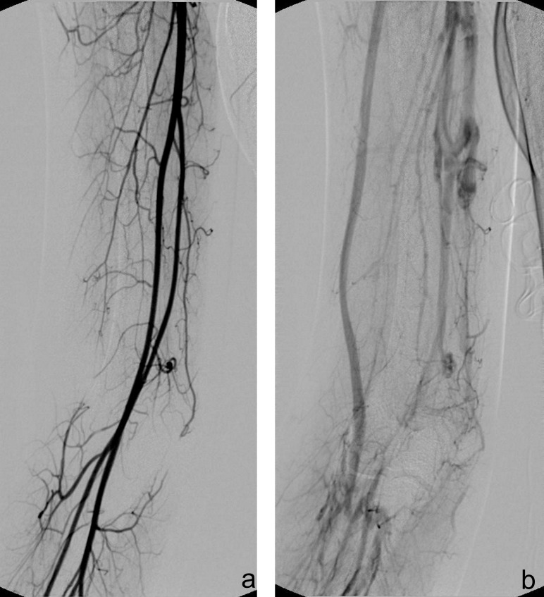
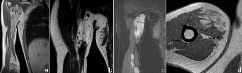
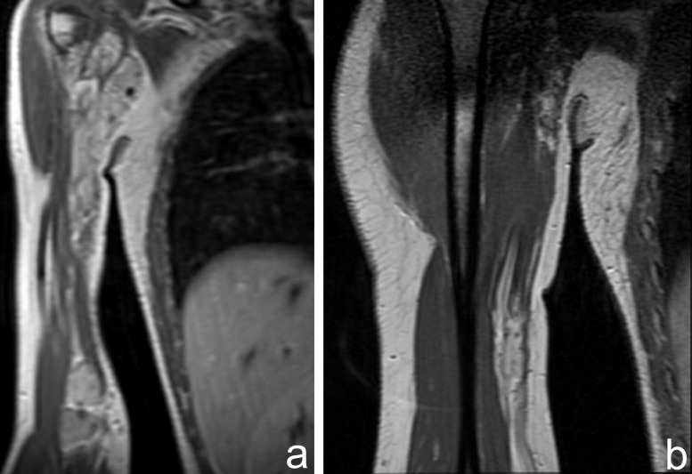
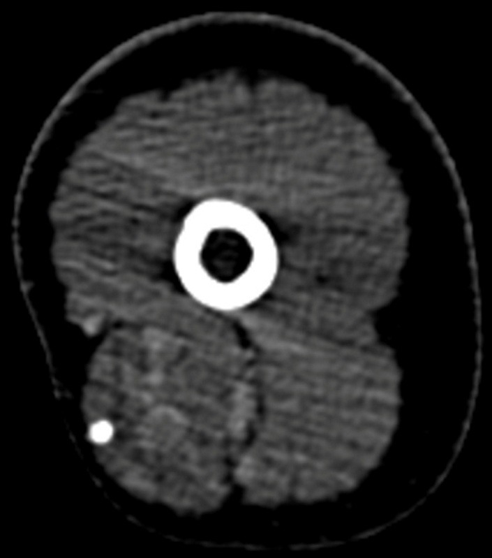
Sinusoidal hemangioma is a rare type of cavernous hemangioma with different clinico-pathological aspects. They are usually localized in the extremities with interest in the subcutaneous layer. The new imaging techniques play an important role in diagnosis, evaluation and follow-up of these types of tumors. We describe the case of a 21-year-old patient, four times operated for a recurrent soft tissue tumor, located intramuscularly in the distal third of the upper limb. Plain X-ray and computer tomography (CT) showed a nonspecific mass with calcification. The MRI (magnetic resonance imaging) exam demonstrated a lobulated heterogeneous signal tumor mass in the biceps brachial muscles, with high signal intensity on T2 weighted images and intermediate signal on T1 weighted images. MRI accurately assessed the extent of the tumor and evaluated the recurrence. MRI imagings combined with contrast-enhanced sequences were used to classify the lesions in low flow vascular disorders. CT angiography with multiplanar reconstructions (MPR), maximum intensity projections (MIP) and volume-rendered reconstructions (VR) was useful in confirming the venous origin of the tumor.

摘要

窦性血管瘤是一种罕见的海绵状血管瘤,具有不同的临床病理特征。它们通常位于四肢,多见于皮下层。新的成像技术在这些类型肿瘤的诊断、评估和随访中发挥着重要作用。我们描述了一名21岁患者的病例,该患者因复发性软组织肿瘤接受了四次手术,肿瘤位于上肢远端三分之一的肌肉内。平片和计算机断层扫描(CT)显示有钙化的非特异性肿块。磁共振成像(MRI)检查显示肱二头肌中有一个分叶状的异质性信号肿瘤肿块,在T2加权图像上呈高信号强度,在T1加权图像上呈中等信号。MRI准确评估了肿瘤的范围并评估了复发情况。结合对比增强序列的MRI成像用于将病变分类为低血流血管疾病。具有多平面重建(MPR)、最大强度投影(MIP)和容积再现重建(VR)的CT血管造影有助于确认肿瘤的静脉起源。

https://cdn.ncbi.nlm.nih.gov/pmc/blobs/cb44/3956096/272bc711f9da/JMedLife-07-46-g001.jpg

相似文献

1
Role of MRI in the diagnosis and evaluation of cavernous hemangioma of the arm.
J Med Life. 2014 Mar 15;7(1):46-50. Epub 2014 Mar 25.
3
Intramuscular cavernous haemangioma of the triceps.
Pan Afr Med J. 2020 Jun 30;36:139. doi: 10.11604/pamj.2020.36.139.23494. eCollection 2020.
6
Computed tomography and magnetic resonance imaging findings of nasal cavity hemangiomas according to histological type.
Korean J Radiol. 2015 May-Jun;16(3):566-74. doi: 10.3348/kjr.2015.16.3.566. Epub 2015 May 13.
7
Evaluation of hemangioma by positron emission tomography: role in a multimodality approach.
J Comput Assist Tomogr. 2003 Jan-Feb;27(1):70-7. doi: 10.1097/00004728-200301000-00013.
8
Magnetic resonance imaging of intramuscular haemangiomas with emphasis on contrast enhancement patterns.
Clin Radiol. 1996 Mar;51(3):198-204. doi: 10.1016/s0009-9260(96)80323-0.
9
Epidural spinal cavernous hemangioma.
Spine (Phila Pa 1976). 1998 May 15;23(10):1136-40. doi: 10.1097/00007632-199805150-00013.
10
CT and MRI manifestations of mediastinal cavernous hemangioma and a review of the literature.
World J Surg Oncol. 2019 Dec 4;17(1):205. doi: 10.1186/s12957-019-1742-1.

引用本文的文献

1
Bleeding cavernous hemangioma of lower lip: a case report.
Ann Med Surg (Lond). 2023 Mar 15;85(5):1867-1869. doi: 10.1097/MS9.0000000000000292. eCollection 2023 May.

本文引用的文献

1
A case of sinusoidal hemangioma with lipoma.
Ann Dermatol. 2011 Oct;23(Suppl 2):S250-3. doi: 10.5021/ad.2011.23.S2.S250. Epub 2011 Oct 31.
2
Vascular anomalies of the upper limb.
Indian J Plast Surg. 2011 May;44(2):276-82. doi: 10.4103/0970-0358.85349.
4
Intramuscular hemangiomas of the extremities: report of six cases.
Int J Dermatol. 2009 Aug;48(8):875-8. doi: 10.1111/j.1365-4632.2008.04033.x.
5
Imaging of peripheral arteries by 16-row multidetector computed tomography angiography: a feasible tool?
Eur J Radiol. 2007 Mar;61(3):528-33. doi: 10.1016/j.ejrad.2006.10.009. Epub 2006 Nov 22.
6
Imaging strategies in intramuscular haemangiomas: an analysis of 20 cases.
Int Orthop. 2007 Aug;31(4):569-75. doi: 10.1007/s00264-006-0229-1. Epub 2006 Oct 5.
7
Multidetector-row CT angiography of upper- and lower-extremity peripheral arteries.
Eur Radiol. 2005 Nov;15 Suppl 4:D3-9. doi: 10.1007/s10406-005-0132-7.
8
Vascular malformations in the extremities: emphasis on MR imaging features that guide treatment options.
Skeletal Radiol. 2006 Mar;35(3):127-37. doi: 10.1007/s00256-005-0057-1. Epub 2006 Jan 27.
9
Circulating level of vascular endothelial growth factor in differentiating hemangioma from vascular malformation patients.
Plast Reconstr Surg. 2005 Jul;116(1):200-4. doi: 10.1097/01.prs.0000170804.80834.5f.
10
Vascular anomalies and the growth of limbs: a review.
J Pediatr Orthop B. 2004 Nov;13(6):349-57. doi: 10.1097/01202412-200411000-00001.

文献AI研究员

20分钟写一篇综述,助力文献阅读效率提升50倍。

立即体验

用中文搜PubMed

大模型驱动的PubMed中文搜索引擎

马上搜索

文档翻译

学术文献翻译模型,支持多种主流文档格式。

立即体验