• 文献检索
  • 文档翻译
  • 深度研究
  • 学术资讯
  • Suppr Zotero 插件Zotero 插件
  • 邀请有礼
  • 套餐&价格
  • 历史记录
应用&插件
Suppr Zotero 插件Zotero 插件浏览器插件Mac 客户端Windows 客户端微信小程序
定价
高级版会员购买积分包购买API积分包
服务
文献检索文档翻译深度研究API 文档MCP 服务
关于我们
关于 Suppr公司介绍联系我们用户协议隐私条款
关注我们

Suppr 超能文献

核心技术专利:CN118964589B侵权必究
粤ICP备2023148730 号-1Suppr @ 2026

文献检索

告别复杂PubMed语法,用中文像聊天一样搜索,搜遍4000万医学文献。AI智能推荐,让科研检索更轻松。

立即免费搜索

文件翻译

保留排版,准确专业,支持PDF/Word/PPT等文件格式,支持 12+语言互译。

免费翻译文档

深度研究

AI帮你快速写综述,25分钟生成高质量综述,智能提取关键信息,辅助科研写作。

立即免费体验

一项使用正面光学相干断层扫描对网状假性玻璃膜疣与脉络膜血管系统进行地形相关性的试点定量研究。

A pilot quantitative study of topographic correlation between reticular pseudodrusen and the choroidal vasculature using en face optical coherence tomography.

作者信息

Grewal Dilraj S, Chou Jonathan, Rollins Stuart D, Fawzi Amani A

机构信息

Department of Ophthalmology, Northwestern University Feinberg School of Medicine, Chicago, Illinois, United States of America.

出版信息

PLoS One. 2014 Mar 21;9(3):e92841. doi: 10.1371/journal.pone.0092841. eCollection 2014.

DOI:10.1371/journal.pone.0092841
PMID:24658298
原文链接:https://pmc.ncbi.nlm.nih.gov/articles/PMC3962468/
Abstract

PURPOSE

To analyze the topographic correlation between reticular pseudodrusen (RPD) visualized on infrared reflectance (IR) and choroidal vasculature using en-face volumetric spectral-domain optical coherence tomography (SD-OCT).

METHODS

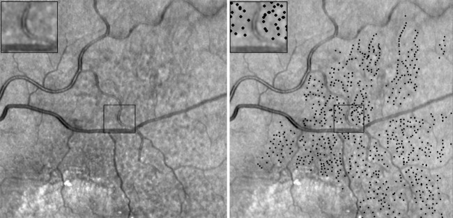
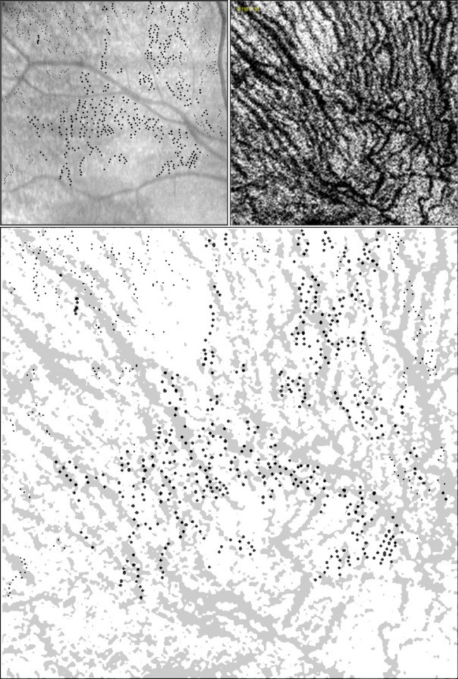
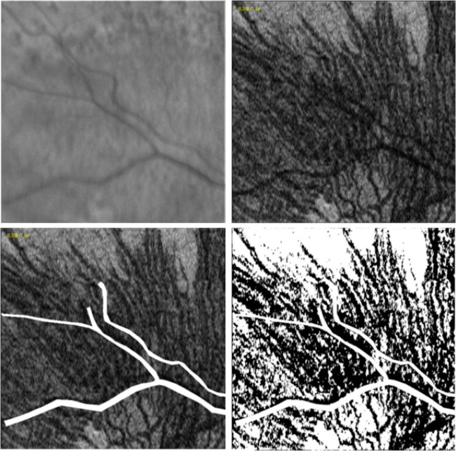
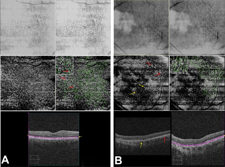
A masked observer marked individual RPD on IR images using ImageJ (NIH, Bethesda, MD). Using the macular volume scan (Cirrus, Carl Zeiss Meditec Inc, Dublin, CA), the RPE slab function was used to generate a C-scan of the most superficial choroidal vasculature. An independent masked grader created a topographic binary map of the choroidal vasculature by thresholding the en-face image, which was overlaid onto the IR map of RPD. For each IR image, ImageJ was used to generate a random set of dots as "control lesions".

RESULTS

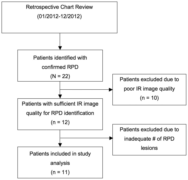
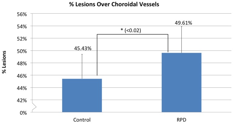
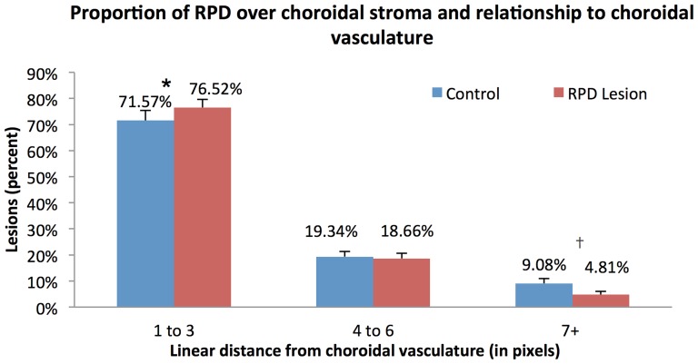
17 eyes of 11 patients (78±13.7 years) with RPD were analyzed. The average number of RPD lesions identified on IR images was 414±71.5, of which 49.6±4.3% were located overlying the choroidal vasculature, compared to 45.4±4.0% in controls (p = 0.014). 50.4±4.3% of lesions overlay the choroidal stroma, of which 76.5±3.1% were ≤3 pixels from the choroidal vessels. The percentage of RPD lesions located within ≤3 pixels from the choroidal vasculature was significantly greater than the percentage located ≥7 pixels away. (p<0.0001). Compared to controls (71.6±3.8%), RPD were more likely to be located ≤3 pixels away from choroidal vessels (p = 0.014). In contrast, control lesions were more likely to be ≥7 pixels away from choroidal vessels than RPD (9.1±1.9% vs. 4.8±1.2%, respectively, p = 0.002).

CONCLUSIONS

Our analysis shows that RPD lesions follow the underlying choroidal vasculature. Approximately half the RPD directly overlay the choroidal vessels and the majority of the remaining lesions were ≤3 pixels (≤30 microns) from the vessel edge, supporting the hypothesis that RPD maybe related to pathologic changes at the choroidal level.

摘要

目的

使用正面容积光谱域光学相干断层扫描(SD-OCT)分析红外反射(IR)下可视化的网状假性玻璃膜疣(RPD)与脉络膜血管系统之间的地形相关性。

方法

一名蒙面观察者使用ImageJ(美国国立卫生研究院,马里兰州贝塞斯达)在IR图像上标记单个RPD。使用黄斑容积扫描(Cirrus,卡尔蔡司医疗技术公司,加利福尼亚州都柏林),RPE平板功能用于生成最浅表脉络膜血管系统的C扫描。一名独立的蒙面分级员通过对正面图像进行阈值处理创建脉络膜血管系统的地形二元图,该图叠加在RPD的IR图上。对于每张IR图像,使用ImageJ生成一组随机点作为“对照病变”。

结果

分析了11例(78±13.7岁)患有RPD的患者的17只眼睛。在IR图像上识别出的RPD病变的平均数量为414±71.5个,其中49.6±4.3%位于脉络膜血管系统上方,而对照组为45.4±4.0%(p = 0.014)。50.4±4.3%的病变覆盖脉络膜基质,其中76.5±3.1%距离脉络膜血管≤3个像素。位于距脉络膜血管系统≤3个像素内的RPD病变百分比显著大于位于≥7个像素外的百分比(p<0.0001)。与对照组(71.6±3.8%)相比,RPD更有可能位于距脉络膜血管≤3个像素处(p = 0.014)。相比之下,对照病变比RPD更有可能距离脉络膜血管≥7个像素(分别为9.1±1.9%和4.8±1.2%,p = 0.002)。

结论

我们的分析表明,RPD病变遵循潜在的脉络膜血管系统。大约一半的RPD直接覆盖脉络膜血管,其余大多数病变距离血管边缘≤3个像素(≤30微米),支持RPD可能与脉络膜水平的病理变化有关的假设。

https://cdn.ncbi.nlm.nih.gov/pmc/blobs/02b4/3962468/3f9548c9318c/pone.0092841.g007.jpg
https://cdn.ncbi.nlm.nih.gov/pmc/blobs/02b4/3962468/d9a58dab0daa/pone.0092841.g001.jpg
https://cdn.ncbi.nlm.nih.gov/pmc/blobs/02b4/3962468/5b546d717aaf/pone.0092841.g002.jpg
https://cdn.ncbi.nlm.nih.gov/pmc/blobs/02b4/3962468/2df2d33f4fa2/pone.0092841.g003.jpg
https://cdn.ncbi.nlm.nih.gov/pmc/blobs/02b4/3962468/1bfca6d4cf0c/pone.0092841.g004.jpg
https://cdn.ncbi.nlm.nih.gov/pmc/blobs/02b4/3962468/1a63ffa03ccd/pone.0092841.g005.jpg
https://cdn.ncbi.nlm.nih.gov/pmc/blobs/02b4/3962468/763d256bc3ca/pone.0092841.g006.jpg
https://cdn.ncbi.nlm.nih.gov/pmc/blobs/02b4/3962468/3f9548c9318c/pone.0092841.g007.jpg

相似文献

1
A pilot quantitative study of topographic correlation between reticular pseudodrusen and the choroidal vasculature using en face optical coherence tomography.一项使用正面光学相干断层扫描对网状假性玻璃膜疣与脉络膜血管系统进行地形相关性的试点定量研究。
PLoS One. 2014 Mar 21;9(3):e92841. doi: 10.1371/journal.pone.0092841. eCollection 2014.
2
RETICULAR PSEUDODRUSEN ON INFRARED IMAGING ARE TOPOGRAPHICALLY DISTINCT FROM SUBRETINAL DRUSENOID DEPOSITS ON EN FACE OPTICAL COHERENCE TOMOGRAPHY.红外成像上的网状假性玻璃膜疣在地形学上与正面光学相干断层扫描上的视网膜下类玻璃膜疣沉积物不同。
Retina. 2015 Dec;35(12):2593-603. doi: 10.1097/IAE.0000000000000666.
3
CHOROIDAL VASCULARITY INDEX AND CHOROIDAL THICKNESS IN EYES WITH RETICULAR PSEUDODRUSEN.伴有网状假性DR 的眼的脉络膜血管指数和脉络膜厚度。
Retina. 2020 Apr;40(4):612-617. doi: 10.1097/IAE.0000000000002667.
4
A pilot study of morphometric analysis of choroidal vasculature in vivo, using en face optical coherence tomography.应用光学相干断层扫描对脉络膜血管进行形态计量分析的初步研究。
PLoS One. 2012;7(11):e48631. doi: 10.1371/journal.pone.0048631. Epub 2012 Nov 26.
5
Macular choroidal thickness and volume of eyes with reticular pseudodrusen using swept-source optical coherence tomography.应用扫频源光学相干断层扫描测量伴网状假性黄斑玻璃膜疣的脉络膜视网膜厚度和体积。
Am J Ophthalmol. 2014 May;157(5):994-1004. doi: 10.1016/j.ajo.2014.01.018. Epub 2014 Jan 31.
6
Localized reticular pseudodrusen and their topographic relation to choroidal watershed zones and changes in choroidal volumes.局限性网状假性色素沉着及其与脉络膜分水岭区的关系和脉络膜容积的变化。
Invest Ophthalmol Vis Sci. 2013 May 7;54(5):3250-7. doi: 10.1167/iovs.13-11923.
7
Widefield En Face Optical Coherence Tomography Imaging of Subretinal Drusenoid Deposits.视网膜下类玻璃膜疣沉积物的广角正面光学相干断层扫描成像
Ophthalmic Surg Lasers Imaging Retina. 2015 May;46(5):550-9. doi: 10.3928/23258160-20150521-06.
8
Image registration and multimodal imaging of reticular pseudodrusen.网状假性小体的图像配准和多模态成像。
Invest Ophthalmol Vis Sci. 2011 Jul 29;52(8):5743-8. doi: 10.1167/iovs.10-6942.
9
Comparison between Widefield En Face Swept-Source OCT and Conventional Multimodal Imaging for the Detection of Reticular Pseudodrusen.广角光相干断层扫描血管成像与传统多模态成像在网状假性黄斑营养不良检测中的比较
Ophthalmology. 2017 Feb;124(2):205-214. doi: 10.1016/j.ophtha.2016.10.009. Epub 2016 Nov 14.
10
Exploring choriocapillaris under reticular pseudodrusen using OCT-Angiography.使用光学相干断层扫描血管造影术探索网状假性玻璃膜疣下的脉络膜毛细血管。
Graefes Arch Clin Exp Ophthalmol. 2016 Nov;254(11):2165-2173. doi: 10.1007/s00417-016-3375-1. Epub 2016 May 18.

引用本文的文献

1
Subretinal Drusenoid Deposits in a Patient With HELLP (Hemolysis, Elevated Liver Enzymes, Low Platelet) Syndrome.一名患有HELLP(溶血、肝酶升高、血小板减少)综合征患者的视网膜下类玻璃膜疣沉积物
Cureus. 2024 Jan 14;16(1):e52239. doi: 10.7759/cureus.52239. eCollection 2024 Jan.
2
Prevalence and determinants of subretinal drusenoid deposits in patients' first-degree relatives.患者一级亲属中视网膜下类脂沉积的患病率及其决定因素。
Graefes Arch Clin Exp Ophthalmol. 2024 Jan;262(1):53-60. doi: 10.1007/s00417-023-06221-y. Epub 2023 Sep 6.
3
Subretinal drusenoid deposits: An update.

本文引用的文献

1
Review of en-face choroidal imaging using spectral-domain optical coherence tomography.使用频域光学相干断层扫描对脉络膜进行表面成像的综述。
Med Hypothesis Discov Innov Ophthalmol. 2013 Fall;2(3):69-73.
2
Phase-variance optical coherence tomography: a technique for noninvasive angiography.相衬光学相干断层扫描:一种无创血管造影技术。
Ophthalmology. 2014 Jan;121(1):180-187. doi: 10.1016/j.ophtha.2013.09.002. Epub 2013 Oct 21.
3
Impact of reticular pseudodrusen on macular function.网状假性色素沉着对黄斑功能的影响。
视网膜下类玻璃膜疣沉积物:最新进展
Taiwan J Ophthalmol. 2022 May 26;12(2):138-146. doi: 10.4103/tjo.tjo_18_22. eCollection 2022 Apr-Jun.
4
Ocular Imaging for Enhancing the Understanding, Assessment, and Management of Age-Related Macular Degeneration.眼部影像学在增强对年龄相关性黄斑变性的理解、评估和管理中的作用。
Adv Exp Med Biol. 2021;1256:33-66. doi: 10.1007/978-3-030-66014-7_2.
5
Subtype-differentiated impacts of subretinal drusenoid deposits on photoreceptors revealed by adaptive optics scanning laser ophthalmoscopy.自适应光学扫描激光检眼镜揭示了脉络膜视网膜下渗出性沉积物对光感受器的亚型分化影响。
Graefes Arch Clin Exp Ophthalmol. 2020 Sep;258(9):1931-1940. doi: 10.1007/s00417-020-04774-w. Epub 2020 Jun 2.
6
Choriocapillaris Vascular Density Changes in Patients with Drusen: Cross-Sectional Study Based on Optical Coherence Tomography Angiography Findings.玻璃膜疣患者脉络膜毛细血管血管密度变化:基于光学相干断层扫描血管造影结果的横断面研究
Ophthalmol Ther. 2018 Jun;7(1):101-107. doi: 10.1007/s40123-018-0119-9. Epub 2018 Jan 30.
7
Intraretinal Correlates of Reticular Pseudodrusen Revealed by Autofluorescence and En Face OCT.通过自发荧光和正面光学相干断层扫描揭示的网状假性玻璃膜疣的视网膜内相关因素
Invest Ophthalmol Vis Sci. 2017 Sep 1;58(11):4769-4777. doi: 10.1167/iovs.17-22338.
8
Choriocapillaris' alterations in the presence of reticular pseudodrusen compared to drusen: study based on OCTA findings.与玻璃膜疣相比,网状假性玻璃膜疣存在时脉络膜毛细血管的改变:基于光学相干断层扫描血管造影(OCTA)结果的研究
Int Ophthalmol. 2018 Oct;38(5):1887-1893. doi: 10.1007/s10792-017-0671-7. Epub 2017 Aug 4.
9
Choroidal Thickness and Choroidal Vessel Density in Nonexudative Age-Related Macular Degeneration Using Swept-Source Optical Coherence Tomography Imaging.使用扫频光学相干断层扫描成像技术测量非渗出性年龄相关性黄斑变性患者的脉络膜厚度和脉络膜血管密度
Invest Ophthalmol Vis Sci. 2016 Nov 1;57(14):6256-6264. doi: 10.1167/iovs.16-20161.
10
Choriocapillaris Nonperfusion is Associated With Poor Visual Acuity in Eyes With Reticular Pseudodrusen.脉络膜毛细血管无灌注与网状玻璃膜疣眼的视力低下有关。
Am J Ophthalmol. 2017 Feb;174:42-55. doi: 10.1016/j.ajo.2016.10.005. Epub 2016 Oct 26.
Retina. 2014 Feb;34(2):321-9. doi: 10.1097/IAE.0b013e3182993df1.
4
Localized reticular pseudodrusen and their topographic relation to choroidal watershed zones and changes in choroidal volumes.局限性网状假性色素沉着及其与脉络膜分水岭区的关系和脉络膜容积的变化。
Invest Ophthalmol Vis Sci. 2013 May 7;54(5):3250-7. doi: 10.1167/iovs.13-11923.
5
Genetic and environmental factors associated with reticular pseudodrusen in age-related macular degeneration.与年龄相关性黄斑变性中网状假性色素沉着相关的遗传和环境因素。
Retina. 2013 May;33(5):998-1004. doi: 10.1097/IAE.0b013e31827b6483.
6
Subretinal drusenoid deposits in non-neovascular age-related macular degeneration: morphology, prevalence, topography, and biogenesis model.非新生血管性年龄相关性黄斑变性的视网膜下类脂沉积:形态、患病率、分布和发生模型。
Retina. 2013 Feb;33(2):265-76. doi: 10.1097/IAE.0b013e31827e25e0.
7
A pilot study of morphometric analysis of choroidal vasculature in vivo, using en face optical coherence tomography.应用光学相干断层扫描对脉络膜血管进行形态计量分析的初步研究。
PLoS One. 2012;7(11):e48631. doi: 10.1371/journal.pone.0048631. Epub 2012 Nov 26.
8
Prevalence and genomic association of reticular pseudodrusen in age-related macular degeneration.年龄相关性黄斑变性中网状假性色素沉着的流行率和基因组关联。
Am J Ophthalmol. 2013 Feb;155(2):260-269.e2. doi: 10.1016/j.ajo.2012.08.011. Epub 2012 Oct 27.
9
NIH Image to ImageJ: 25 years of image analysis.NIH 图像到 ImageJ:25 年的图像分析。
Nat Methods. 2012 Jul;9(7):671-5. doi: 10.1038/nmeth.2089.
10
Choroidal changes associated with reticular pseudodrusen.与网状假性小体相关的脉络膜改变。
Invest Ophthalmol Vis Sci. 2012 Mar 9;53(3):1258-63. doi: 10.1167/iovs.11-8907.