MI-Lab, Department of Medical Imaging, St. Olavs University Hospital Trondheim, Norway ; Erwin L. Hahn Institute of Magnetic Resonance Imaging, University Duisburg-Essen Essen, Germany.
Erwin L. Hahn Institute of Magnetic Resonance Imaging, University Duisburg-Essen Essen, Germany ; Centre for Cognitive Neuroimaging, Donders Institute for Brain, Cognition and Behaviour, Radboud University Nijmegen Nijmegen, Netherlands.
Front Neurosci. 2014 Mar 13;8:49. doi: 10.3389/fnins.2014.00049. eCollection 2014.
To compare the BOLD fMRI signal characteristics at in the cortex and on the pial surface for a non-balanced steady-state free precession sequence (nb-SSFP) at 7 T.
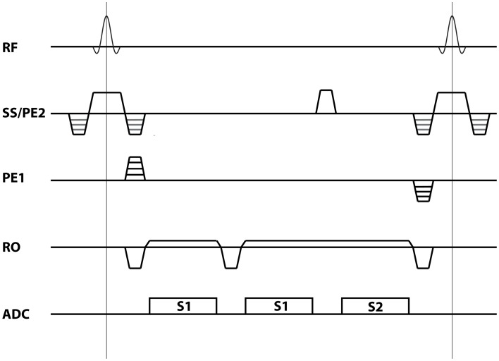
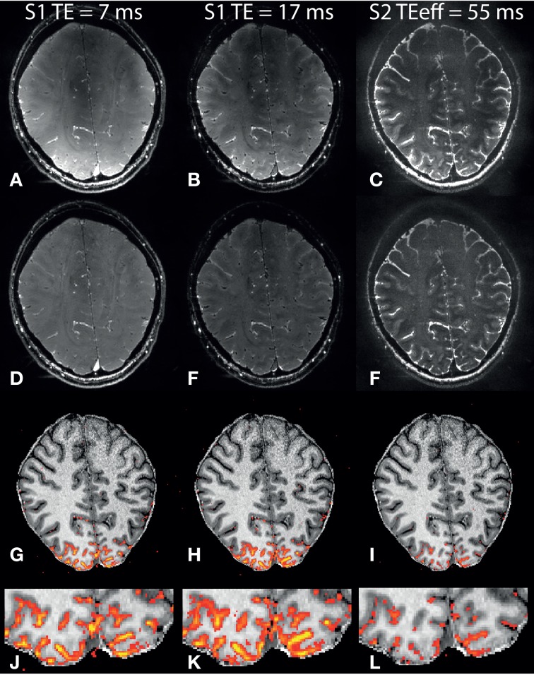
A multi-echo nb-SSFP sequence was used for high resolution fMRI at 7 T. Two S1 (S(+)) echoes at different echo times were acquired together with an S2 (S(-)) echo. The primary visual cortex (V1) was examined using a reversing checkerboard paradigm at an isotropic resolution of 0.75 mm, with 35 volumes acquired and a total scan time of 27 min.
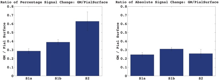
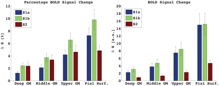
Significant activation was observed in all subjects for all three acquired echoes. For the S1 signal at the longer TE, the activation induced signal change was about 4% in the cortex and 10% at the cortical surface, while for S2 the corresponding values were 3 and 5%.
For both S1 and S2 data, the BOLD signal peaks at the pial surface. The large pial surface signal change in S2 may be caused by dynamic averaging around post-capillary vessels embedded within CSF. This is made possible by the long diffusion times of the pathways contributing to the S2 signal and the relatively high diffusion coefficient of CSF. The results indicate that S2-SSFP might not be a suited alternative to spin-echo for high-resolution fMRI at 7 T.
比较在 7T 下非平衡稳态自由进动序列(nb-SSFP)皮层和脑表面的 BOLD fMRI 信号特征。
使用多回波 nb-SSFP 序列在 7T 下进行高分辨率 fMRI。同时采集两个不同回波时间的 S1(S(+))回波和一个 S2(S(-))回波。使用反转棋盘格范式在各向同性分辨率为 0.75mm 下检查初级视觉皮层(V1),采集 35 个容积,总扫描时间为 27 分钟。
所有受试者在所有三个采集回波中均观察到显著激活。对于较长 TE 的 S1 信号,皮层内的激活诱导信号变化约为 4%,皮层表面为 10%,而对于 S2,相应的值分别为 3%和 5%。
对于 S1 和 S2 数据,BOLD 信号均在脑表面达到峰值。S2 中脑表面信号的较大变化可能是由于嵌入在 CSF 中的毛细血管后血管周围的动态平均所致。这是由于 S2 信号的路径具有较长的扩散时间和相对较高的 CSF 扩散系数而成为可能。结果表明,S2-SSFP 可能不是 7T 高分辨率 fMRI 中自旋回波的合适替代方法。