Department of Otolaryngology - Head and Neck Surgery, Johns Hopkins University School of Medicine , Baltimore, MD , USA.
Department of Neurology, Johns Hopkins University School of Medicine , Baltimore, MD , USA.
Front Neurol. 2014 Mar 13;5:28. doi: 10.3389/fneur.2014.00028. eCollection 2014.
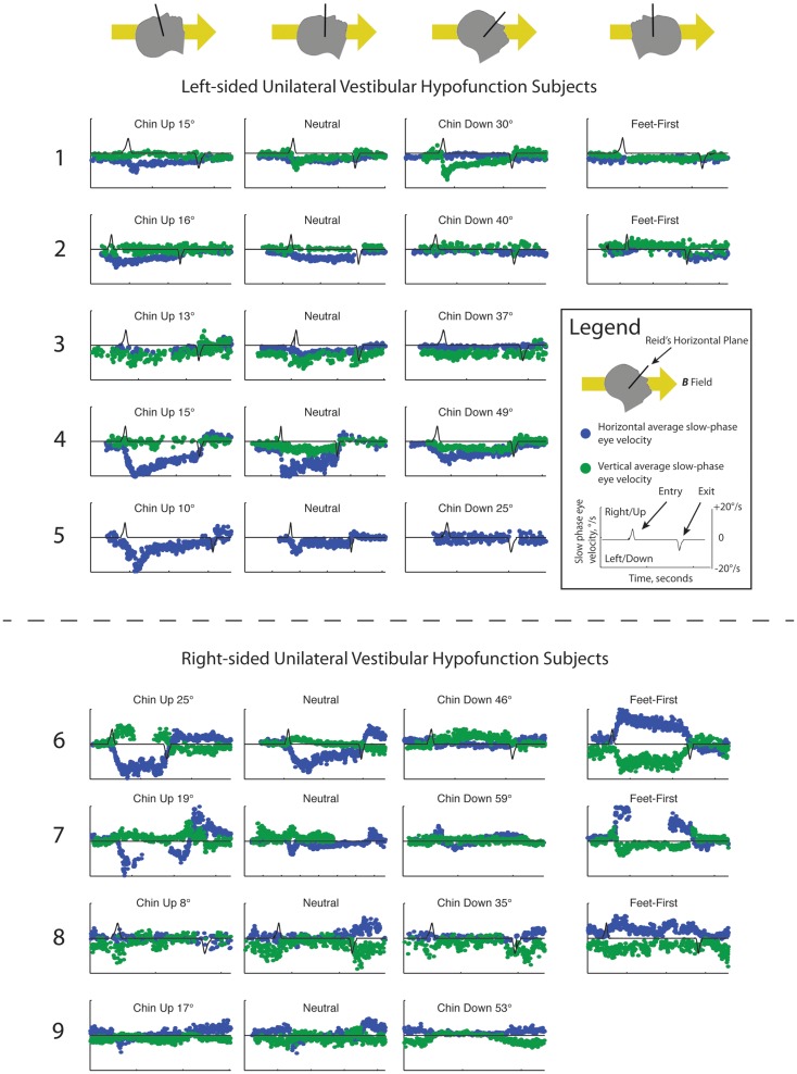
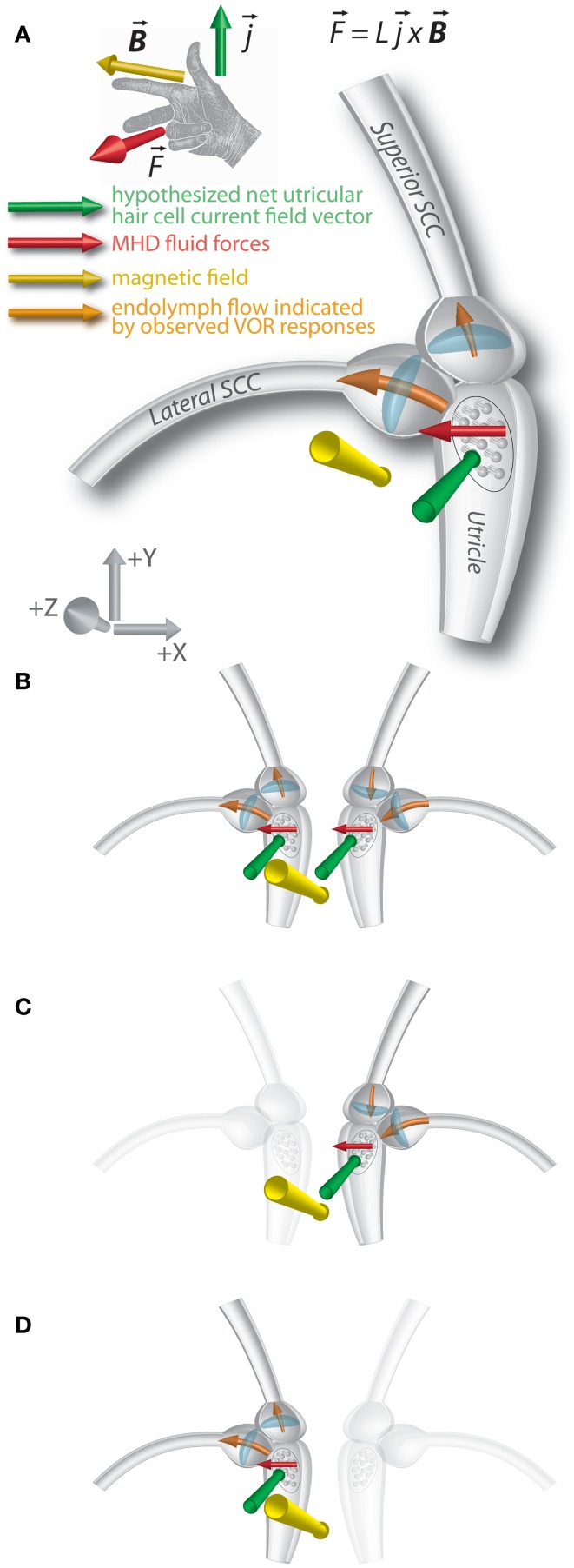
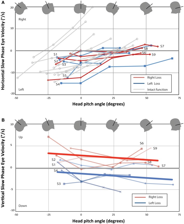
We recently discovered that static magnetic fields from high-strength MRI machines induce nystagmus in all normal humans, and that a magneto-hydrodynamic Lorentz force, derived from ionic currents in the endolymph and pushing on the cupula, best explains this effect. Individuals with no labyrinthine function have no nystagmus. The influence of magnetic vestibular stimulation (MVS) in individuals with unilateral deficits in labyrinthine function is unknown and may provide insight into the mechanism of MVS. These individuals should experience MVS, but with a different pattern of nystagmus consistent with their unilateral deficit in labyrinthine function. We recorded eye movements in the static magnetic field of a 7 T MRI machine in nine individuals with unilateral labyrinthine hypofunction, as determined by head impulse testing and vestibular-evoked myogenic potentials (VEMP). Eye movements were recorded using infrared video-oculography. Static head positions were varied in pitch with the body supine, and slow-phase eye velocity (SPV) was assessed. All subjects exhibited predominantly horizontal nystagmus after entering the magnet head-first, lying supine. The SPV direction reversed when entering feet-first. Pitching chin-to-chest caused subjects to reach a null point for horizontal SPV. Right unilateral vestibular hypofunction (UVH) subjects developed slow-phase-up nystagmus and left UVH subjects, slow-phase-down nystagmus. Vertical and torsional components were consistent with superior semicircular canal excitation or inhibition, respectively, of the intact ear. These findings provide compelling support for the hypothesis that MVS is a result of a Lorentz force and suggest that the function of individual structures within the labyrinth can be assessed with MVS. As a novel method of comfortable and sustained labyrinthine stimulation, MVS can provide new insights into vestibular physiology and pathophysiology.
我们最近发现,高强度 MRI 机器产生的静态磁场会引起所有正常人出现眼球震颤,而源自内淋巴中离子电流并推动镫骨的磁流体动力学洛伦兹力能够很好地解释这一现象。没有迷路功能的个体则不会出现眼球震颤。磁前庭刺激(MVS)对单侧迷路功能不全个体的影响尚不清楚,这可能有助于深入了解 MVS 的机制。这些个体应该会经历 MVS,但会出现与单侧迷路功能不全相一致的不同眼球震颤模式。我们通过头脉冲测试和前庭诱发肌源性电位(VEMP)确定了 9 名单侧迷路功能低下个体的单侧迷路功能低下,并在 7T MRI 机器的静态磁场中记录了他们的眼球运动。使用红外视频眼动记录眼球运动。在仰卧体位下,头部以矢状面方向改变静态头位,评估慢相眼速度(SPV)。所有受试者在头先进磁体、仰卧位时,都会先出现以水平方向为主的眼球震颤。当脚先进磁体时,SPV 方向会反转。低头使颏部贴近胸部会使受试者达到水平 SPV 的零位。单侧右侧(UVH)前庭功能低下受试者会出现慢相向上的眼球震颤,而左侧 UVH 受试者则出现慢相向下的眼球震颤。垂直和扭转成分分别与对侧完整耳的上半规管兴奋或抑制相一致。这些发现为 MVS 是洛伦兹力的结果这一假说提供了有力支持,并表明可以通过 MVS 评估迷路内单个结构的功能。作为一种舒适且持续的迷路刺激的新方法,MVS 可以为前庭生理学和病理生理学提供新的见解。