Raz Eytan, Loh John P, Saba Luca, Omari Mirza, Herbert Joseph, Lui Yvonne, Kister Ilya
Department of Radiology, New York University School of Medicine, 660 First Avenue, New York, NY 10026, USA.
Department of Radiology, Azienda Ospedaliero Universitaria, di Cagliari Polo di Monserrato, SS 554 Monserrato, 09045 Cagliari, Italy.
Mult Scler Int. 2014;2014:986923. doi: 10.1155/2014/986923. Epub 2014 Feb 9.
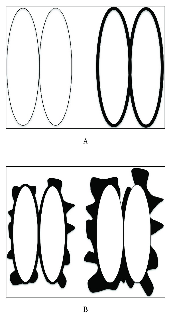
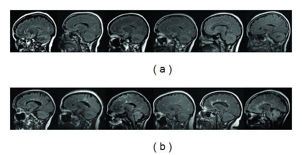
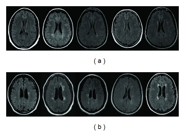
Objective. To compare periventricular lesions in multiple sclerosis (MS) and neuromyelitis optica spectrum disorders (NMOsd). Materials and Methods. Sagittal and axial fluid attenuated inversion recovery (FLAIR) sequences of 20 NMOsd and 40 group frequency-matched MS patients were evaluated by two neuroradiologists. On axial FLAIR, periventricular area was characterized as free of lesions/smooth-bordered ("type A") or jagged-bordered ("type B") pattern. On sagittal FLAIR, the images were evaluated for presence of "Dawson's fingers." Results. Type A pattern was observed in 80% of NMOsd patients by Reader 1 and 85% by Reader 2 but only in 5% MS patients by either Reader. Type B was seen in 15% NMOsd patients by Reader 1 and 20% by Reader 2 and in 95% MS patients by either Reader. Dawson's fingers were observed in no NMOsd patients by Reader 1 and 5% by Reader 2. In MS, Dawson's fingers were seen in 92.5% patients by Reader 1 and 77.5% by Reader 2. The differences in periventricular patterns and Dawson's finger detection between NMOsd and MS were highly significant (P < 0.001). Conclusions. Dawson's fingers and "jagged-bordered" periventricular hyperintensities are typical of MS and almost never seen in NMOsd, which suggests a practical method for differentiating the two diseases.
目的。比较多发性硬化(MS)和视神经脊髓炎谱系障碍(NMOsd)中的脑室周围病变。材料与方法。由两名神经放射科医生对20例NMOsd患者和40例频率匹配的MS患者的矢状位和轴位液体衰减反转恢复(FLAIR)序列进行评估。在轴位FLAIR上,脑室周围区域的特征为无病变/边界光滑(“A型”)或边界参差不齐(“B型”)模式。在矢状位FLAIR上,评估图像是否存在“道森指”。结果。读者1观察到80%的NMOsd患者为A型模式,读者2观察到85%;而MS患者中,两位读者观察到的A型模式均仅为5%。读者1观察到15%的NMOsd患者为B型,读者2观察到20%;而MS患者中,两位读者观察到的B型均为95%。读者1观察到无NMOsd患者存在道森指,读者2观察到5%有。在MS中,读者1观察到92.5%的患者有道森指,读者2观察到77.5%。NMOsd和MS在脑室周围模式和道森指检测方面的差异具有高度显著性(P < 0.001)。结论。道森指和“边界参差不齐”的脑室周围高信号是MS的典型表现,在NMOsd中几乎从未见过,这提示了一种区分这两种疾病的实用方法。