• 文献检索
  • 文档翻译
  • 深度研究
  • 学术资讯
  • Suppr Zotero 插件Zotero 插件
  • 邀请有礼
  • 套餐&价格
  • 历史记录
应用&插件
Suppr Zotero 插件Zotero 插件浏览器插件Mac 客户端Windows 客户端微信小程序
定价
高级版会员购买积分包购买API积分包
服务
文献检索文档翻译深度研究API 文档MCP 服务
关于我们
关于 Suppr公司介绍联系我们用户协议隐私条款
关注我们

Suppr 超能文献

核心技术专利:CN118964589B侵权必究
粤ICP备2023148730 号-1Suppr @ 2026

文献检索

告别复杂PubMed语法,用中文像聊天一样搜索,搜遍4000万医学文献。AI智能推荐,让科研检索更轻松。

立即免费搜索

文件翻译

保留排版,准确专业,支持PDF/Word/PPT等文件格式,支持 12+语言互译。

免费翻译文档

深度研究

AI帮你快速写综述,25分钟生成高质量综述,智能提取关键信息,辅助科研写作。

立即免费体验

小鼠创伤性脑损伤后神经胶质细胞群体的皮质增殖的时间模式。

Temporal patterns of cortical proliferation of glial cell populations after traumatic brain injury in mice.

机构信息

‡Developmental Neurobiology Section, Division of Intramural Research, National Heart, Lung, and Blood Institute, National Institutes of Health, Bethesda, MD, U.S.A.

出版信息

ASN Neuro. 2014 May 8;6(3):159-70. doi: 10.1042/AN20130034.

DOI:10.1042/AN20130034
PMID:24670035
原文链接:https://pmc.ncbi.nlm.nih.gov/articles/PMC4013687/
Abstract

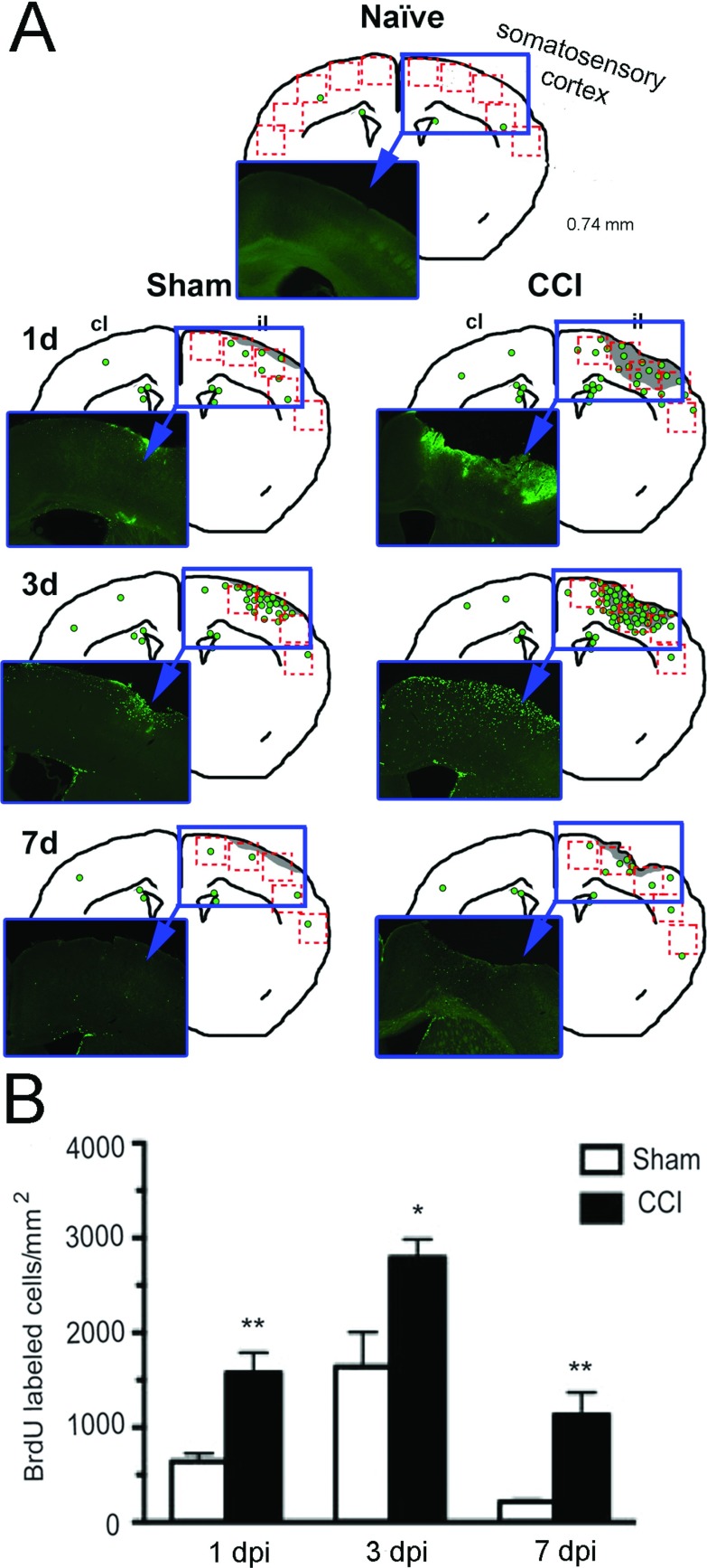
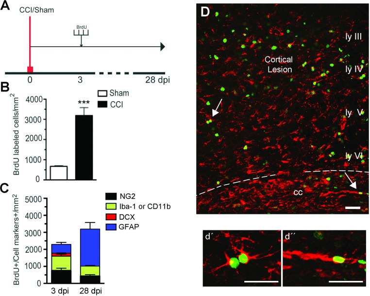
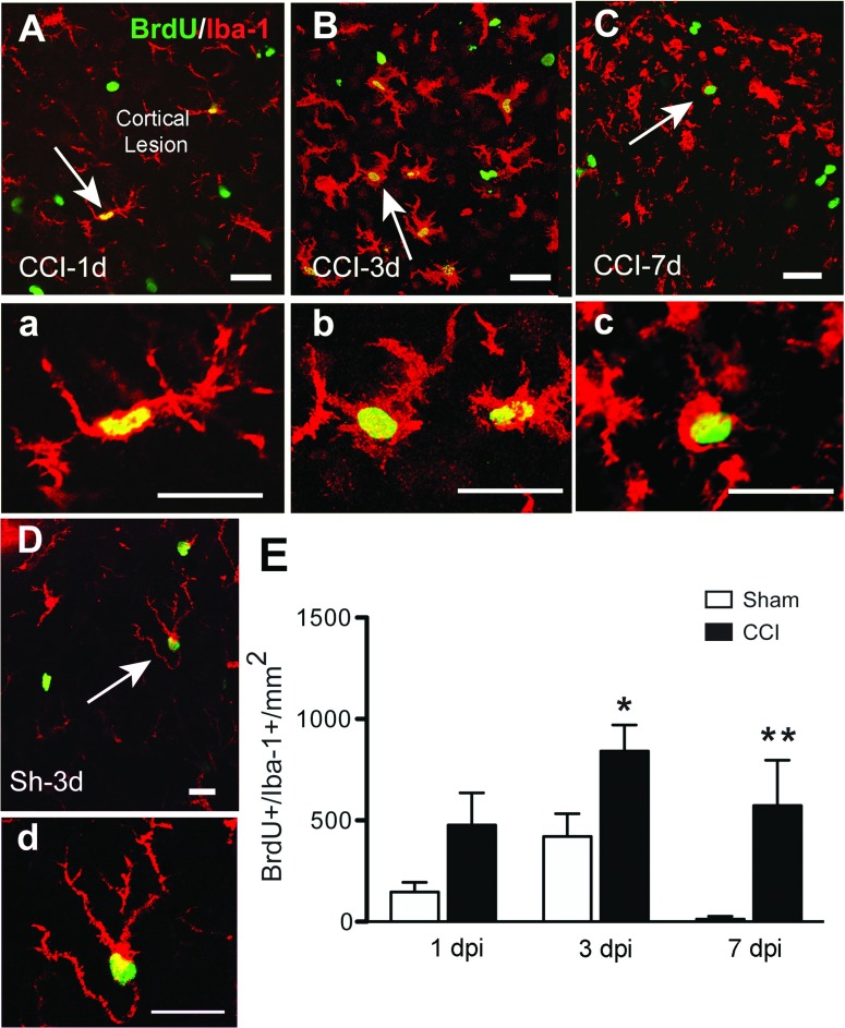
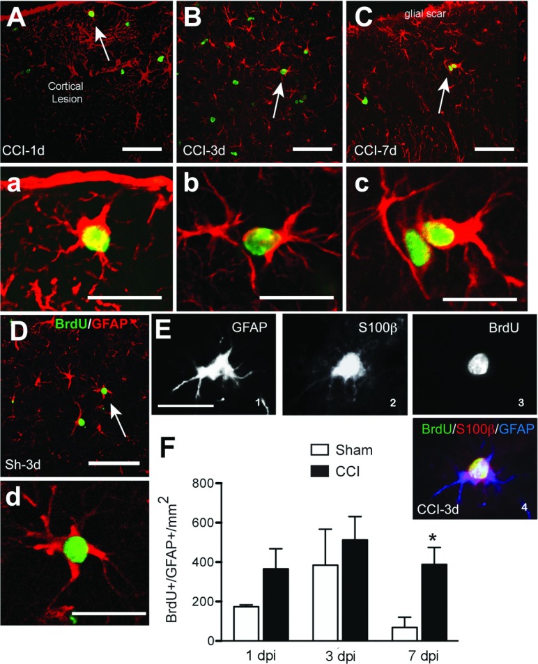
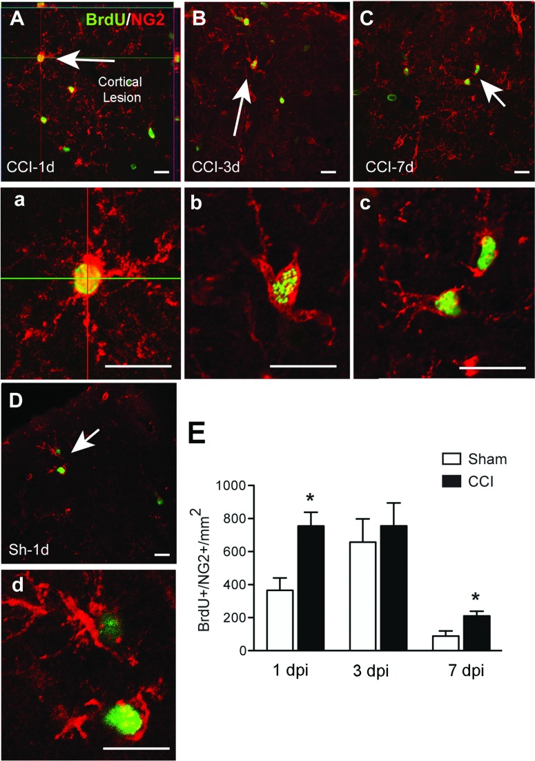
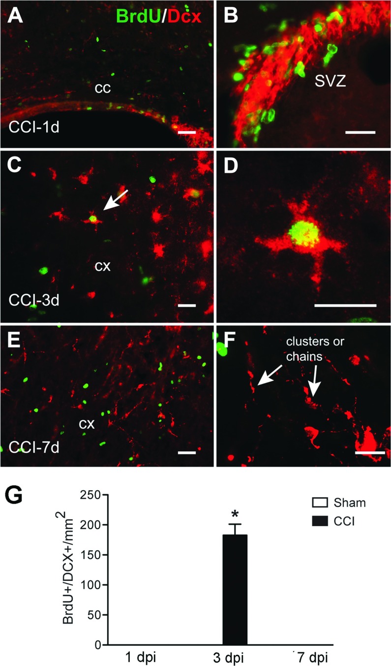
TBI (traumatic brain injury) triggers an inflammatory cascade, gliosis and cell proliferation following cell death in the pericontusional area and surrounding the site of injury. In order to better understand the proliferative response following CCI (controlled cortical impact) injury, we systematically analyzed the phenotype of dividing cells at several time points post-lesion. C57BL/6 mice were subjected to mild to moderate CCI over the left sensory motor cortex. At different time points following injury, mice were injected with BrdU (bromodeoxyuridine) four times at 3-h intervals and then killed. The greatest number of proliferating cells in the pericontusional region was detected at 3 dpi (days post-injury). At 1 dpi, NG2+ cells were the most proliferative population, and at 3 and 7 dpi the Iba-1+ microglial cells were proliferating more. A smaller, but significant number of GFAP+ (glial fibrillary acidic protein) astrocytes proliferated at all three time points. Interestingly, at 3 dpi we found a small number of proliferating neuroblasts [DCX+ (doublecortin)] in the injured cortex. To determine the cell fate of proliferative cells, mice were injected four times with BrdU at 3 dpi and killed at 28 dpi. Approximately 70% of proliferative cells observed at 28 dpi were GFAP+ astrocytes. In conclusion, our data suggest that the specific glial cell types respond differentially to injury, suggesting that each cell type responds to a specific pattern of growth factor stimulation at each time point after injury.

摘要

脑创伤(TBI)会在挫伤区及其周围的损伤部位引发细胞死亡后的炎症级联反应、神经胶质增生和细胞增殖。为了更好地理解 CCI(皮质控制冲击)损伤后的增殖反应,我们系统地分析了损伤后几个时间点分裂细胞的表型。将 C57BL/6 小鼠的左感觉运动皮层置于轻度至中度 CCI 之下。在损伤后不同的时间点,将 BrdU(溴脱氧尿苷)注射到小鼠体内,每 3 小时注射一次,共注射四次,然后杀死它们。在损伤后 3 天(dpi)检测到挫伤区周围增殖细胞的数量最多。在 1 dpi 时,NG2+细胞是最具增殖性的群体,而在 3 和 7 dpi 时,Iba-1+小胶质细胞增殖更多。一小部分但具有统计学意义的 GFAP+(胶质纤维酸性蛋白)星形胶质细胞在三个时间点都有增殖。有趣的是,在 3 dpi 时,我们在损伤皮层中发现了一小部分增殖的神经前体细胞[DCX+(双皮质素)]。为了确定增殖细胞的命运,在 3 dpi 时将 BrdU 注射四次,然后在 28 dpi 时杀死小鼠。在 28 dpi 时观察到的增殖细胞中,约有 70%是 GFAP+星形胶质细胞。总之,我们的数据表明,特定的神经胶质细胞类型对损伤的反应不同,这表明每种细胞类型在损伤后每个时间点对特定的生长因子刺激模式都有反应。

https://cdn.ncbi.nlm.nih.gov/pmc/blobs/9747/4013687/6da4b91caa62/an2013-0034i006.jpg
https://cdn.ncbi.nlm.nih.gov/pmc/blobs/9747/4013687/065931b59fa1/an2013-0034i001.jpg
https://cdn.ncbi.nlm.nih.gov/pmc/blobs/9747/4013687/3f6080a94419/an2013-0034i002.jpg
https://cdn.ncbi.nlm.nih.gov/pmc/blobs/9747/4013687/b23953a29add/an2013-0034i003.jpg
https://cdn.ncbi.nlm.nih.gov/pmc/blobs/9747/4013687/1aa483f53245/an2013-0034i004.jpg
https://cdn.ncbi.nlm.nih.gov/pmc/blobs/9747/4013687/99fd7fcd37a2/an2013-0034i005.jpg
https://cdn.ncbi.nlm.nih.gov/pmc/blobs/9747/4013687/6da4b91caa62/an2013-0034i006.jpg

相似文献

1
Temporal patterns of cortical proliferation of glial cell populations after traumatic brain injury in mice.小鼠创伤性脑损伤后神经胶质细胞群体的皮质增殖的时间模式。
ASN Neuro. 2014 May 8;6(3):159-70. doi: 10.1042/AN20130034.
2
Aging increases microglial proliferation, delays cell migration, and decreases cortical neurogenesis after focal cerebral ischemia.衰老会增加小胶质细胞增殖,延迟细胞迁移,并减少局灶性脑缺血后的皮质神经发生。
J Neuroinflammation. 2015 May 10;12:87. doi: 10.1186/s12974-015-0314-8.
3
Cortical endogenic neural regeneration of adult rat after traumatic brain injury.成年大鼠创伤性脑损伤后的皮质内源性神经再生。
PLoS One. 2013 Jul 29;8(7):e70306. doi: 10.1371/journal.pone.0070306. Print 2013.
4
Granulocyte-colony stimulating factor promotes brain repair following traumatic brain injury by recruitment of microglia and increasing neurotrophic factor expression.粒细胞集落刺激因子通过募集小胶质细胞和增加神经营养因子表达来促进创伤性脑损伤后的脑修复。
Restor Neurol Neurosci. 2016 Feb 24;34(3):415-31. doi: 10.3233/RNN-150607.
5
Astrocytes and oligodendrocytes can be generated from NG2+ progenitors after acute brain injury: intracellular localization of oligodendrocyte transcription factor 2 is associated with their fate choice.急性脑损伤后,NG2+祖细胞可生成星形胶质细胞和少突胶质细胞:少突胶质细胞转录因子2的细胞内定位与其命运选择相关。
Eur J Neurosci. 2009 May;29(9):1853-69. doi: 10.1111/j.1460-9568.2009.06736.x. Epub 2009 Apr 28.
6
Lack of NG2 exacerbates neurological outcome and modulates glial responses after traumatic brain injury.NG2 缺失加剧创伤性脑损伤后的神经功能预后,并调节神经胶质反应。
Glia. 2016 Apr;64(4):507-23. doi: 10.1002/glia.22944. Epub 2015 Dec 6.
7
Developmental and post-injury cortical gliogenesis: a genetic fate-mapping study with Nestin-CreER mice.发育性和损伤后皮质神经胶质生成:利用巢蛋白-CreER小鼠进行的遗传命运图谱研究。
Glia. 2009 Aug 1;57(10):1115-29. doi: 10.1002/glia.20835.
8
Osteopontin is indispensable for activation of astrocytes in injured mouse brain and primary culture.骨桥蛋白对于损伤小鼠脑和原代培养中星形胶质细胞的激活不可或缺。
Neurol Res. 2018 Dec;40(12):1071-1079. doi: 10.1080/01616412.2018.1517995. Epub 2018 Sep 22.
9
Alterations in hippocampal neurogenesis following traumatic brain injury in mice.小鼠创伤性脑损伤后海马神经发生的改变。
Exp Neurol. 2006 Nov;202(1):189-99. doi: 10.1016/j.expneurol.2006.05.034. Epub 2006 Jul 28.
10
Cellular proliferation and migration following a controlled cortical impact in the mouse.小鼠控制性皮质撞击后的细胞增殖与迁移
Brain Res. 2005 Aug 16;1053(1-2):38-53. doi: 10.1016/j.brainres.2005.06.042.

引用本文的文献

1
Longitudinal Brain Structural, Neurochemical, and Behavioral Changes Following Traumatic Brain Injury in Immature Rat Brain with ALCAR Treatment.用ALCAR治疗的未成熟大鼠脑创伤性脑损伤后的纵向脑结构、神经化学和行为变化
Neurochem Res. 2025 Jul 30;50(4):256. doi: 10.1007/s11064-025-04494-9.
2
Chronic starvation induces microglial cell depletion in an activity-based anorexia model.在基于活动的厌食症模型中,慢性饥饿会导致小胶质细胞耗竭。
Sci Rep. 2025 Apr 23;15(1):14132. doi: 10.1038/s41598-025-98237-z.
3
Temporal characterisation and electrophysiological implications of TBI-induced serine/threonine kinase activity in mouse cortex.

本文引用的文献

1
Microglia: Multiple roles in surveillance, circuit shaping, and response to injury.小胶质细胞:在监视、回路形成和对损伤的反应中的多种作用。
Neurology. 2013 Sep 17;81(12):1079-88. doi: 10.1212/WNL.0b013e3182a4a577. Epub 2013 Aug 14.
2
The Time Course of Gene Expression during Reactive Gliosis in the Optic Nerve.视神经反应性胶质增生过程中基因表达的时间进程。
PLoS One. 2013 Jun 27;8(6):e67094. doi: 10.1371/journal.pone.0067094. Print 2013.
3
Controlled cortical impact and craniotomy induce strikingly similar profiles of inflammatory gene expression, but with distinct kinetics.
创伤性脑损伤诱导的小鼠皮质丝氨酸/苏氨酸激酶活性的时间特征及电生理意义
Cell Mol Life Sci. 2025 Mar 5;82(1):102. doi: 10.1007/s00018-025-05638-4.
4
Post-injury treatment with 7,8-dihydroxyflavone attenuates white matter pathology in aged mice following focal traumatic brain injury.创伤后用7,8-二羟基黄酮治疗可减轻老年小鼠局灶性创伤性脑损伤后的白质病变。
Neurotherapeutics. 2025 Jan;22(1):e00472. doi: 10.1016/j.neurot.2024.e00472. Epub 2024 Oct 20.
5
Astrocytes, reactive astrogliosis, and glial scar formation in traumatic brain injury.创伤性脑损伤中的星形胶质细胞、反应性星形胶质增生和胶质瘢痕形成。
Neural Regen Res. 2025 Apr 1;20(4):973-989. doi: 10.4103/NRR.NRR-D-23-02091. Epub 2024 May 17.
6
Traumatic brain injury promotes neurogenesis at the cost of astrogliogenesis in the adult hippocampus of male mice.创伤性脑损伤以星形胶质细胞发生为代价促进成年雄性小鼠海马中的神经发生。
Nat Commun. 2024 Jun 18;15(1):5222. doi: 10.1038/s41467-024-49299-6.
7
A primary culture method for the easy, efficient, and effective acquisition of oligodendrocyte lineage cells from neonatal rodent brains.一种从新生啮齿动物大脑中轻松、高效且有效地获取少突胶质细胞谱系细胞的原代培养方法。
Heliyon. 2024 Apr 12;10(8):e29359. doi: 10.1016/j.heliyon.2024.e29359. eCollection 2024 Apr 30.
8
Glial cells react to closed head injury in a distinct and spatiotemporally orchestrated manner.神经胶质细胞对闭合性颅脑损伤的反应具有独特的、时空协调的方式。
Sci Rep. 2024 Jan 30;14(1):2441. doi: 10.1038/s41598-024-52337-4.
9
Microglial histone deacetylase 2 is dispensable for functional and histological outcomes in a mouse model of traumatic brain injury.小胶质细胞组蛋白去乙酰化酶 2 在创伤性脑损伤小鼠模型中的功能和组织学结果中不是必需的。
J Cereb Blood Flow Metab. 2024 May;44(5):817-835. doi: 10.1177/0271678X231197173. Epub 2023 Dec 9.
10
Intranasal Leukemia Inhibitory Factor Attenuates Gliosis and Axonal Injury and Improves Sensorimotor Function After a Mild Pediatric Traumatic Brain Injury.鼻内注射白血病抑制因子可减轻小儿轻度创伤性脑损伤后的胶质增生和轴突损伤,并改善感觉运动功能。
Neurotrauma Rep. 2023 Apr 11;4(1):236-250. doi: 10.1089/neur.2021.0075. eCollection 2023.
控制性皮质撞击和开颅术诱导出极为相似的炎症基因表达谱,但具有不同的动力学特征。
Front Neurol. 2012 Oct 31;3:155. doi: 10.3389/fneur.2012.00155. eCollection 2012.
4
Candesartan, an angiotensin II AT₁-receptor blocker and PPAR-γ agonist, reduces lesion volume and improves motor and memory function after traumatic brain injury in mice.坎地沙坦,一种血管紧张素 II AT₁受体阻滞剂和 PPAR-γ 激动剂,可减少创伤性脑损伤后小鼠的损伤体积,并改善运动和记忆功能。
Neuropsychopharmacology. 2012 Dec;37(13):2817-29. doi: 10.1038/npp.2012.152. Epub 2012 Aug 15.
5
The effect of injury severity on behavior: a phenotypic study of cognitive and emotional deficits after mild, moderate, and severe controlled cortical impact injury in mice.损伤严重程度对行为的影响:在轻度、中度和重度控制性皮质撞击损伤后对认知和情感缺陷的表型研究。
J Neurotrauma. 2012 Sep;29(13):2283-96. doi: 10.1089/neu.2012.2456. Epub 2012 Aug 3.
6
Alterations in sulfated chondroitin glycosaminoglycans following controlled cortical impact injury in mice.在小鼠的控制性皮质撞击损伤后,硫酸软骨素糖胺聚糖的改变。
J Comp Neurol. 2012 Oct 15;520(15):3295-313. doi: 10.1002/cne.23156.
7
Microglia and neuronal cell death.小胶质细胞与神经元细胞死亡。
Neuron Glia Biol. 2011 Feb;7(1):25-40. doi: 10.1017/S1740925X12000014. Epub 2012 Mar 1.
8
Role of the lesion scar in the response to damage and repair of the central nervous system.病灶疤痕在中枢神经系统损伤和修复中的作用。
Cell Tissue Res. 2012 Jul;349(1):169-80. doi: 10.1007/s00441-012-1336-5. Epub 2012 Feb 25.
9
Leptomeningeal-derived doublecortin-expressing cells in poststroke brain.脑卒后脑膜来源的双皮质素阳性细胞。
Stem Cells Dev. 2012 Sep 1;21(13):2350-4. doi: 10.1089/scd.2011.0657. Epub 2012 Apr 2.
10
Genetic deletion of cdc42 reveals a crucial role for astrocyte recruitment to the injury site in vitro and in vivo.遗传敲除 cdc42 揭示了星形胶质细胞在体外和体内向损伤部位募集的关键作用。
J Neurosci. 2011 Aug 31;31(35):12471-82. doi: 10.1523/JNEUROSCI.2696-11.2011.