Gao Jie, Wang He, Liu Yuan, Li Ying-Yu, Chen Can, Liu Liang-Ming, Wu Ya-Min, Li Sen, Yang Ce
State Key Laboratory of Trauma, Burns and Combined Injury, Research Institute of Surgery, Daping Hospital, Third Military Medical University, Chongqing, China (mainland).
Med Sci Monit. 2014 Mar 27;20:499-512. doi: 10.12659/MSM.890589.
People who experience traumatic events have an increased risk of post-traumatic stress disorder (PTSD). However, PTSD-related pathological changes in the hippocampus and prefrontal cortex remain poorly understood.
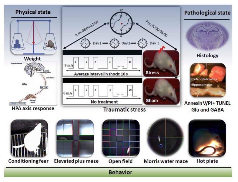
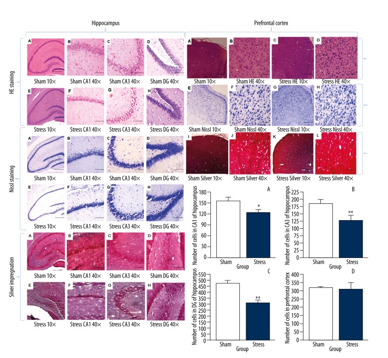
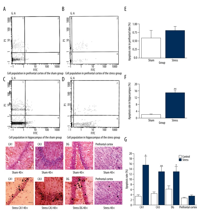
We investigated the effect of a PTSD-like animal model induced by severe stress. The experimental rats received 20 inescapable electric foot shocks in an enclosed box for a total of 6 times in 3 days. The physiological state (body weight and plasma corticosterone concentrations), emotion, cognitive behavior, brain morphology, apoptosis, and balance of gamma-aminobutyric acid (GABA) and glutamate in the hippocampus and prefrontal cortex were observed. Cell damages were examined with histological staining (HE, Nissl, and silver impregnation), while apoptosis was analyzed with flow cytometry using an Annexin V and propidium iodide (PI) binding and terminal deoxynucleotidyl transferase mediated-dUTP nick end labeling (TUNEL) method.
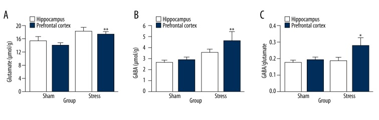
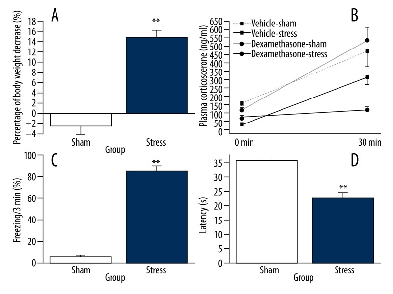
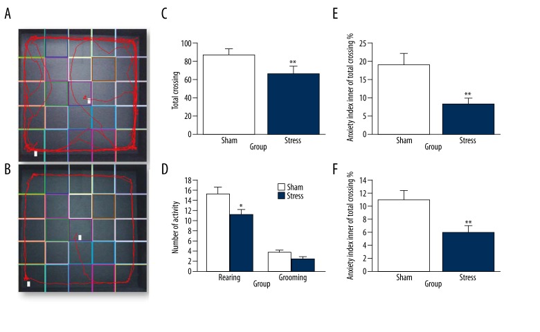
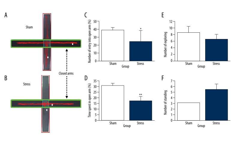
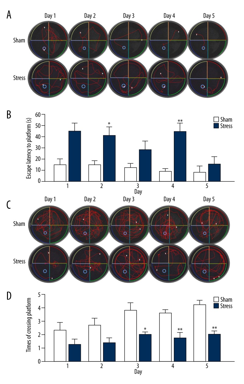
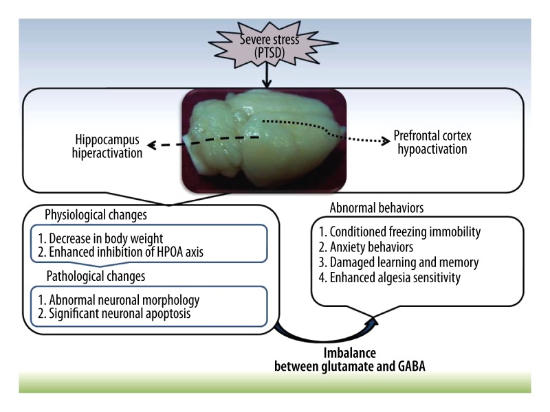
In comparison with the sham litter-mates, the stressed rats showed decreased body weight, inhibition of hypothalamic-pituitary-adrenal (HPA) axis activation, increase in freezing response to trauma reminder, hypoactivity and anxiety-like behaviors in elevated plus maze and open field test, poor learning in Morris water maze, and shortened latency in hot-plate test. There were significant damages in the hippocampus but not in the prefrontal cortex. Imbalance between glutamate and GABA was more evident in the hippocampus than in the prefrontal cortex.
These results suggest that neuronal apoptosis in the hippocampus after severe traumatic stress is related to the imbalance between glutamate and GABA. Such modifications may resemble the profound changes observed in PTSD patients.
经历创伤性事件的人患创伤后应激障碍(PTSD)的风险增加。然而,海马体和前额叶皮质中与PTSD相关的病理变化仍知之甚少。
我们研究了由严重应激诱导的PTSD样动物模型的影响。实验大鼠在封闭箱中接受20次不可逃避的足部电击,3天内共6次。观察其生理状态(体重和血浆皮质酮浓度)、情绪、认知行为、脑形态、细胞凋亡以及海马体和前额叶皮质中γ-氨基丁酸(GABA)和谷氨酸的平衡。用组织学染色(苏木精-伊红染色、尼氏染色和银染)检查细胞损伤,同时使用膜联蛋白V和碘化丙啶(PI)结合以及末端脱氧核苷酸转移酶介导的dUTP缺口末端标记(TUNEL)方法通过流式细胞术分析细胞凋亡。
与假手术同窝仔相比,应激大鼠体重下降,下丘脑-垂体-肾上腺(HPA)轴激活受到抑制,对创伤提示的僵住反应增加,在高架十字迷宫和旷场试验中活动减少和出现焦虑样行为,在莫里斯水迷宫中学习能力差,在热板试验中潜伏期缩短。海马体有明显损伤,但前额叶皮质没有。海马体中谷氨酸和GABA之间的失衡比前额叶皮质更明显。
这些结果表明,严重创伤应激后海马体中的神经元凋亡与谷氨酸和GABA之间的失衡有关。这种改变可能类似于在PTSD患者中观察到的深刻变化。