Chen Christine, Deng Meihong, Sun Qian, Loughran Patricia, Billiar Timothy R, Scott Melanie J
Department of Surgery, University of Pittsburgh, NW607 MUH, 3459 Fifth Avenue, Pittsburgh, PA 15213, USA.
Department of Surgery, University of Pittsburgh, NW607 MUH, 3459 Fifth Avenue, Pittsburgh, PA 15213, USA ; Department of Pathology, University of Pittsburgh, Pittsburgh, PA 15213, USA.
Biomed Res Int. 2014;2014:267350. doi: 10.1155/2014/267350. Epub 2014 Feb 10.
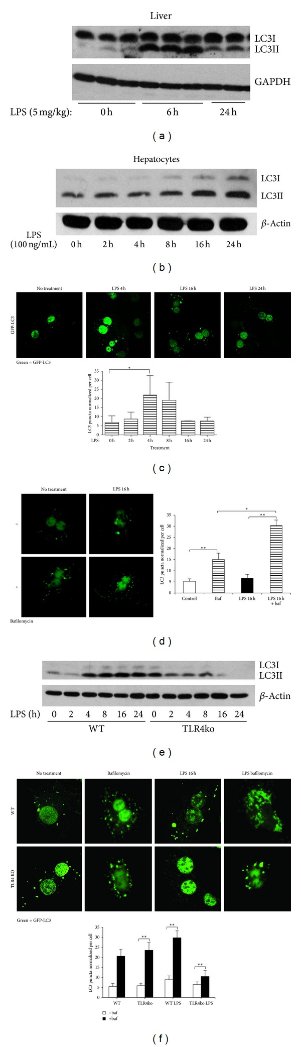
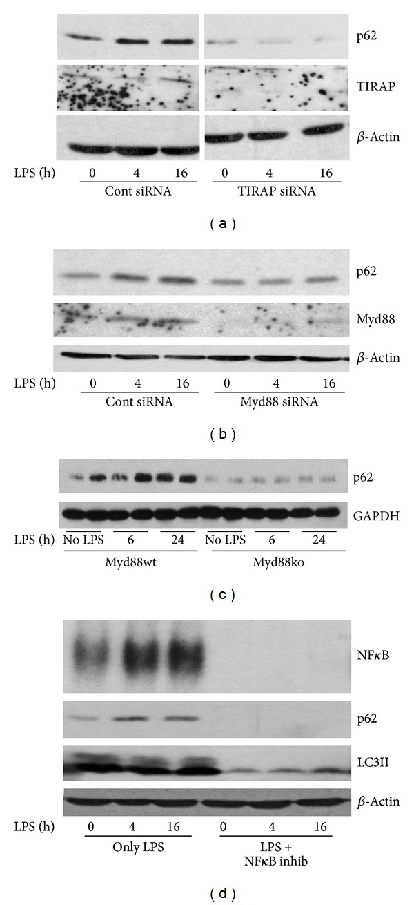
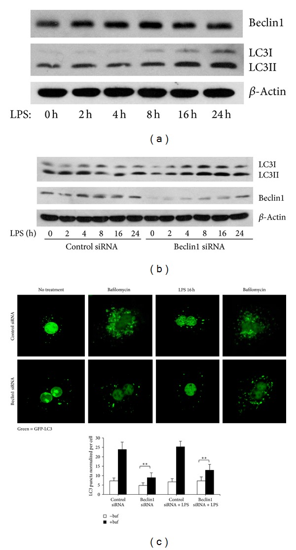
Impairment of autophagy has been associated with liver injury. TLR4-stimulation by LPS upregulates autophagy in hepatocytes, although the signaling pathways involved remain elusive. The objective of this study was to determine the signaling pathway leading to LPS-stimulated autophagy in hepatocytes. Cell lysates from livers of wild type (WT; C57BL/6) mice given LPS (5 mg/kg-IP) and hepatocytes from WT, TLR4ko, and MyD88ko mice treated with LPS (100 ng/mL) up to 24 h were collected. LC3II, p62/SQSTM1, Nrf2, and beclin1 levels were determined by immunoblot, immunofluorescence, and qPCR. Autophagy-like activation was measured by GFP-LC3-puncta formation and LC3II-expression. Beclin1, Nrf2, p62, MyD88, and TIRAP were knocked-down using siRNA. LC3II-expression increased in both liver and hepatocytes after LPS and was dependent on TLR4. Beclin1 expression did not increase after LPS in hepatocytes and beclin1-knockdown did not affect LC3II levels. In hepatocytes given LPS, expression of p62 increased and p62 colocalized with LC3. p62-knockdown prevented LC3II puncta formation. LPS-induced LC3II/p62-puncta also required MyD88/TIRAP signaling and localization of both Nrf2 and NF κ B transcription factors to the nucleus to upregulate p62-expression. Therefore, TLR4-activation by LPS in hepatocytes induces a p62-mediated, not beclin1-mediated, autophagy-like clearance pathway that is hepatoprotective by clearing aggregate-prone or misfolded proteins from the cytosol and preserving energy homeostasis under stress.
自噬功能受损与肝损伤有关。脂多糖(LPS)刺激Toll样受体4(TLR4)可上调肝细胞中的自噬,尽管其中涉及的信号通路仍不清楚。本研究的目的是确定导致LPS刺激肝细胞自噬的信号通路。收集野生型(WT;C57BL/6)小鼠腹腔注射LPS(5mg/kg)后肝脏的细胞裂解物,以及WT、TLR4基因敲除(TLR4ko)和髓样分化因子88基因敲除(MyD88ko)小鼠的肝细胞,这些肝细胞用LPS(100ng/mL)处理长达24小时。通过免疫印迹、免疫荧光和定量聚合酶链反应(qPCR)测定微管相关蛋白1轻链3-II(LC3II)、p62/ sequestosome 1(p62/SQSTM1)、核因子E2相关因子2(Nrf2)和自噬相关蛋白1(beclin1)的水平。通过绿色荧光蛋白-LC3斑点形成和LC3II表达来测量自噬样激活。使用小干扰RNA(siRNA)敲低beclin1、Nrf2、p62、MyD88和TIRAP。LPS处理后,肝脏和肝细胞中的LC3II表达均增加,且依赖于TLR4。LPS处理后,肝细胞中的beclin1表达未增加,beclin1敲低也不影响LC3II水平。在给予LPS的肝细胞中,p62表达增加,且p62与LC3共定位。p62敲低可阻止LC3II斑点形成。LPS诱导的LC3II/p62斑点还需要MyD88/TIRAP信号传导以及Nrf2和核因子κB(NFκB)转录因子定位于细胞核以上调p62表达。因此,LPS激活肝细胞中的TLR4可诱导一种由p62介导而非beclin1介导的自噬样清除途径,该途径通过从细胞质中清除易于聚集或错误折叠的蛋白质并在应激状态下维持能量稳态来发挥肝脏保护作用。