• 文献检索
  • 文档翻译
  • 深度研究
  • 学术资讯
  • Suppr Zotero 插件Zotero 插件
  • 邀请有礼
  • 套餐&价格
  • 历史记录
应用&插件
Suppr Zotero 插件Zotero 插件浏览器插件Mac 客户端Windows 客户端微信小程序
定价
高级版会员购买积分包购买API积分包
服务
文献检索文档翻译深度研究API 文档MCP 服务
关于我们
关于 Suppr公司介绍联系我们用户协议隐私条款
关注我们

Suppr 超能文献

核心技术专利:CN118964589B侵权必究
粤ICP备2023148730 号-1Suppr @ 2026

文献检索

告别复杂PubMed语法,用中文像聊天一样搜索,搜遍4000万医学文献。AI智能推荐,让科研检索更轻松。

立即免费搜索

文件翻译

保留排版,准确专业,支持PDF/Word/PPT等文件格式,支持 12+语言互译。

免费翻译文档

深度研究

AI帮你快速写综述,25分钟生成高质量综述,智能提取关键信息,辅助科研写作。

立即免费体验

用于腭裂闭合的牵引成骨:有限元分析。

Distraction osteogenesis for cleft palate closure: A finite element analysis.

作者信息

Ghasemianpour Majid, Ehsani Sara, Tahmasbi Soodeh, Bayat Mohammad, Ghorbanpour Maedeh, Safavi Seyed Mohammadreza, Mirhashemi Fatemeh Sadat

机构信息

Dental Research center, Research Institute of Dental Sciences, Shahid Beheshti University of Medical Sciences, Tehran, Iran.

Department of Orthodontics, Shahid Beheshti University of Medical Sciences, Tehran, Iran.

出版信息

Dent Res J (Isfahan). 2014 Jan;11(1):92-9.

PMID:24688567
原文链接:https://pmc.ncbi.nlm.nih.gov/articles/PMC3955322/
Abstract

BACKGROUND

Current methods of closure of the cleft palate result in the formation of scars and impairment of growth. Distraction osteogenesis (DO) might be an effective means to repair or at least reduce the size of wide clefts. This study investigates the biomechanical aspects of this process.

MATERIALS AND METHODS

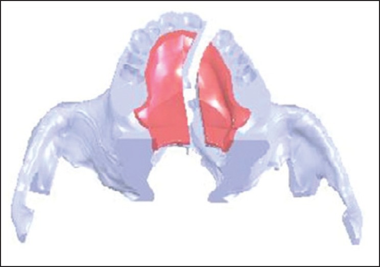
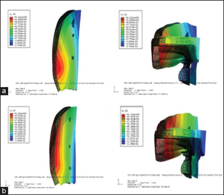
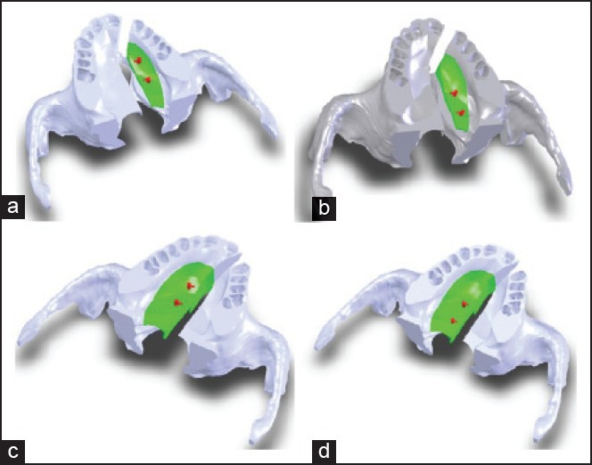
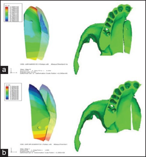
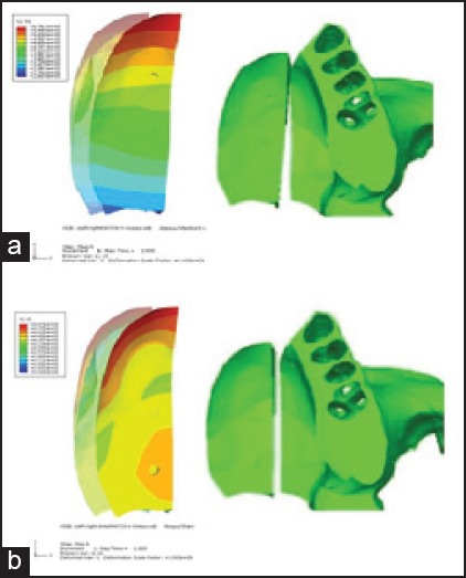
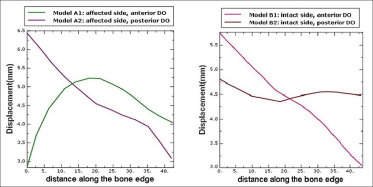
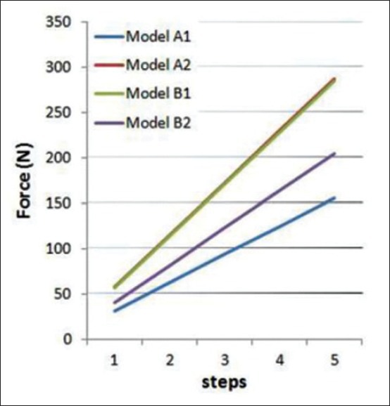
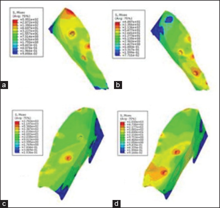
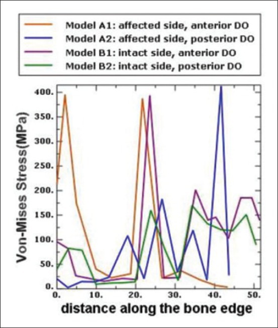
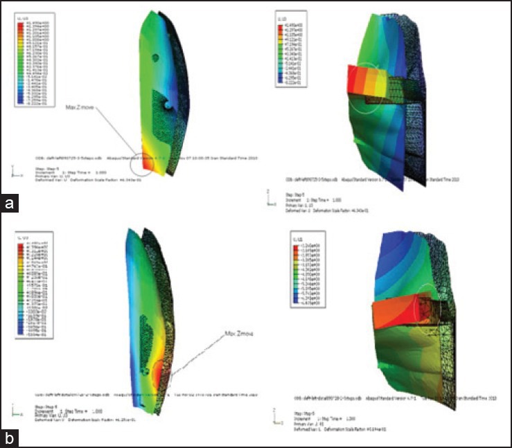
DO simulation was applied to reduce the size of a unilateral hard palate cleft on a three-dimensional (3D) model of the maxilla. For the position of osteotomy lines, two different models were assumed, with the osteotomy line on the affected side in model A and on the intact side in model B. In each model, DO screws were placed on two different positions, anteriorly (models A1 and B1) and posteriorly (models A2 and B2). Displacement pattern of the bony island in each of the four models, reaction forces at DO locations, and von Mises stress were estimated. Mesh generation and data processing were carried out in the 3D finite element analysis package (ABAQUS V6.7-1; Simulia Corp., Providence, RI, USA).

RESULTS

In model B2, the island moved almost evenly, assuring a more complete closure of the cleft. The most uniform stress distribution was found in model B1.

CONCLUSION

The results suggest that the best positions for the DO screw and the osteotomy line for closure of the cleft palate are posteriorly and on the intact side, respectively.

摘要

背景

目前腭裂修复方法会导致瘢痕形成和生长障碍。牵张成骨术(DO)可能是修复或至少减小宽大腭裂尺寸的有效方法。本研究探讨该过程的生物力学方面。

材料与方法

在三维(3D)上颌骨模型上应用DO模拟来减小单侧硬腭裂的尺寸。对于截骨线位置,假设了两种不同模型,模型A中截骨线位于患侧,模型B中截骨线位于健侧。在每个模型中,DO螺钉放置在两个不同位置,前方(模型A1和B1)和后方(模型A2和B2)。估计了四个模型中每个模型的骨岛位移模式、DO位置处的反作用力和冯·米塞斯应力。在3D有限元分析软件包(ABAQUS V6.7 - 1;美国罗德岛州普罗维登斯市Simulia公司)中进行网格生成和数据处理。

结果

在模型B2中,骨岛移动几乎均匀,确保腭裂更完全闭合。在模型B1中发现应力分布最均匀。

结论

结果表明,用于腭裂闭合的DO螺钉和截骨线的最佳位置分别是后方和健侧。

https://cdn.ncbi.nlm.nih.gov/pmc/blobs/2a8b/3955322/431cee43cfa3/DRJ-11-92-g012.jpg
https://cdn.ncbi.nlm.nih.gov/pmc/blobs/2a8b/3955322/c369b9a8ad65/DRJ-11-92-g001.jpg
https://cdn.ncbi.nlm.nih.gov/pmc/blobs/2a8b/3955322/5fded6bb2872/DRJ-11-92-g002.jpg
https://cdn.ncbi.nlm.nih.gov/pmc/blobs/2a8b/3955322/41e5875131b7/DRJ-11-92-g004.jpg
https://cdn.ncbi.nlm.nih.gov/pmc/blobs/2a8b/3955322/3bb7c442f076/DRJ-11-92-g005.jpg
https://cdn.ncbi.nlm.nih.gov/pmc/blobs/2a8b/3955322/5a7de00efa7a/DRJ-11-92-g006.jpg
https://cdn.ncbi.nlm.nih.gov/pmc/blobs/2a8b/3955322/7a2aaec3031e/DRJ-11-92-g008.jpg
https://cdn.ncbi.nlm.nih.gov/pmc/blobs/2a8b/3955322/7c1ac52cc7c7/DRJ-11-92-g009.jpg
https://cdn.ncbi.nlm.nih.gov/pmc/blobs/2a8b/3955322/b20700736e9b/DRJ-11-92-g010.jpg
https://cdn.ncbi.nlm.nih.gov/pmc/blobs/2a8b/3955322/589705049550/DRJ-11-92-g011.jpg
https://cdn.ncbi.nlm.nih.gov/pmc/blobs/2a8b/3955322/431cee43cfa3/DRJ-11-92-g012.jpg

相似文献

1
Distraction osteogenesis for cleft palate closure: A finite element analysis.用于腭裂闭合的牵引成骨:有限元分析。
Dent Res J (Isfahan). 2014 Jan;11(1):92-9.
2
[Three-dimensional finite element analysis for external midface distraction after different osteotomy in patients with cleft lip and palate].[唇腭裂患者不同截骨术后面中份牵张成骨的三维有限元分析]
Zhonghua Zheng Xing Wai Ke Za Zhi. 2010 Jul;26(4):248-51.
3
Sutural expansion osteogenesis for management of the bony-tissue defect in cleft palate repair: experimental studies in dogs.用于腭裂修复中骨组织缺损处理的缝线扩张成骨:犬实验研究
Plast Reconstr Surg. 2000 May;105(6):2012-25; discussion 2026-7. doi: 10.1097/00006534-200005000-00016.
4
Maxillary distraction osteogenesis versus orthognathic surgery for cleft lip and palate patients.唇腭裂患者的上颌骨牵张成骨术与正颌外科手术对比
Cochrane Database Syst Rev. 2018 Aug 10;8(8):CD010403. doi: 10.1002/14651858.CD010403.pub3.
5
[Sutural distraction osteogenesis for primary cleft palate repair: a preliminary clinical report].[用于原发性腭裂修复的缝牵引成骨:初步临床报告]
Zhonghua Zheng Xing Wai Ke Za Zhi. 2003 Jul;19(4):261-4.
6
Distraction osteogenesis for bony repair of cleft palate by using persistent elastic force: experimental study in dogs.利用持续弹力进行腭裂骨修复的牵张成骨:犬实验研究
Cleft Palate Craniofac J. 2005 May;42(3):231-8. doi: 10.1597/03-098.1.
7
[Biomechanical study of internal midface distraction after different types of maxillary osteotomy in patients with cleft lip and palate].[唇腭裂患者不同类型上颌骨截骨术后面中部内牵张成骨的生物力学研究]
Zhonghua Zheng Xing Wai Ke Za Zhi. 2009 Sep;25(5):368-72.
8
Distraction osteogenesis for cleft palate closure in a canine model.犬模型中用于腭裂闭合的牵引成骨术。
Arch Facial Plast Surg. 2005 Nov-Dec;7(6):398-404. doi: 10.1001/archfaci.7.6.398.
9
[Biomechanical effect of 2 anchorages on maxillary protraction with cleft lip and palate: a finite element analysis].[两种支抗对唇腭裂患者上颌前牵引的生物力学影响:有限元分析]
Shanghai Kou Qiang Yi Xue. 2017 Feb;26(1):1-6.
10
[Application of distraction osteogenesis in severe maxillary hypoplasia secondary to cleft palate].牵张成骨术在腭裂继发严重上颌骨发育不全中的应用
Zhonghua Yi Xue Za Zhi. 2002 May 25;82(10):699-702.

引用本文的文献

1
3D Printed Models of Cleft Palate Pathology for Surgical Education.用于外科教育的腭裂病理学3D打印模型
Plast Reconstr Surg Glob Open. 2016 Sep 27;4(9):e1029. doi: 10.1097/GOX.0000000000001029. eCollection 2016 Sep.
2
Biomechanical effects of maxillary expansion on a patient with cleft palate: A finite element analysis.上颌扩弓对腭裂患者的生物力学影响:有限元分析
Am J Orthod Dentofacial Orthop. 2016 Aug;150(2):313-23. doi: 10.1016/j.ajodo.2015.12.029.

本文引用的文献

1
Alveolar cleft closure by osseodistraction: pitfalls and troubleshooting.骨牵引成骨技术用于牙槽嵴裂修复:陷阱与处理方法
J Craniofac Surg. 2012 Mar;23(2):e72-5. doi: 10.1097/SCS.0b013e31824685d3.
2
Biomechanical response of the maxillofacial skeleton to transpalatal orthopedic force in a unilateral palatal cleft.单侧腭裂患者腭中缝骨皮质切开术后上颌骨复合体的生物力学反应。
Angle Orthod. 2011 May;81(3):503-9. doi: 10.2319/070110-367.1. Epub 2011 Feb 7.
3
Biomechanical analysis of rapid maxillary expansion in the UCLP patient.单侧完全性唇腭裂患者快速上颌扩弓的生物力学分析
Med Eng Phys. 2009 Apr;31(3):409-17. doi: 10.1016/j.medengphy.2008.06.011. Epub 2008 Aug 29.
4
A new approach to repairing cleft palate and acquired palatal defects with distraction osteogenesis.一种利用牵张成骨修复腭裂和后天性腭部缺损的新方法。
Int J Oral Maxillofac Surg. 2006 Aug;35(8):718-26. doi: 10.1016/j.ijom.2006.03.010. Epub 2006 May 9.
5
Distraction osteogenesis for cleft palate closure in a canine model.犬模型中用于腭裂闭合的牵引成骨术。
Arch Facial Plast Surg. 2005 Nov-Dec;7(6):398-404. doi: 10.1001/archfaci.7.6.398.
6
Segment distraction to reduce a wide alveolar cleft before alveolar bone grafting.在牙槽骨植骨前进行节段性牵引以缩小较宽的牙槽裂。
Cleft Palate Craniofac J. 2003 Nov;40(6):561-5. doi: 10.1597/1545-1569_2003_040_0561_sdtraw_2.0.co_2.
7
Mandibular distraction in neonates: a strategy to avoid tracheostomy.新生儿下颌骨牵张成骨:一种避免气管切开术的策略。
Plast Reconstr Surg. 2002 Mar;109(3):896-904; discussion 905-6. doi: 10.1097/00006534-200203000-00011.
8
Incidence of cleft palate fistula: an institutional experience with two-stage palatal repair.腭裂瘘的发生率:两阶段腭裂修复术的机构经验
Plast Reconstr Surg. 2001 Nov;108(6):1515-8. doi: 10.1097/00006534-200111000-00011.
9
Closure of vertical alveolar bone defects with guided horizontal distraction osteogenesis: an experimental study in pigs and first clinical results.引导性水平骨牵张成骨术闭合垂直牙槽骨缺损:猪的实验研究及初步临床结果
J Craniomaxillofac Surg. 2001 Oct;29(5):249-53. doi: 10.1054/jcms.2001.0240.
10
Sutural expansion osteogenesis for management of the bony-tissue defect in cleft palate repair: experimental studies in dogs.用于腭裂修复中骨组织缺损处理的缝线扩张成骨:犬实验研究
Plast Reconstr Surg. 2000 May;105(6):2012-25; discussion 2026-7. doi: 10.1097/00006534-200005000-00016.