Lioufas Peter A, Quayle Michelle R, Leong James C, McMenamin Paul G
Monash Health, Monash University, Melbourne, Australia; Centre for Human Anatomy Education, Department of Anatomy & Developmental Biology, School of Biomedical Sciences, Faculty of Medicine, Nursing and Health Sciences, Monash University, Clayton, Australia; and Plastics & Reconstructive Surgery, Department of Surgery, Monash Health, Monash University, Melbourne, Australia.
Plast Reconstr Surg Glob Open. 2016 Sep 27;4(9):e1029. doi: 10.1097/GOX.0000000000001029. eCollection 2016 Sep.
To explore the potential viability and limitations of 3D printed models of children with cleft palate deformity.
The advantages of 3D printed replicas of normal anatomical specimens have previously been described. The creation of 3D prints displaying patient-specific anatomical pathology for surgical planning and interventions is an emerging field. Here we explored the possibility of taking rare pediatric radiographic data sets to create 3D prints for surgical education.
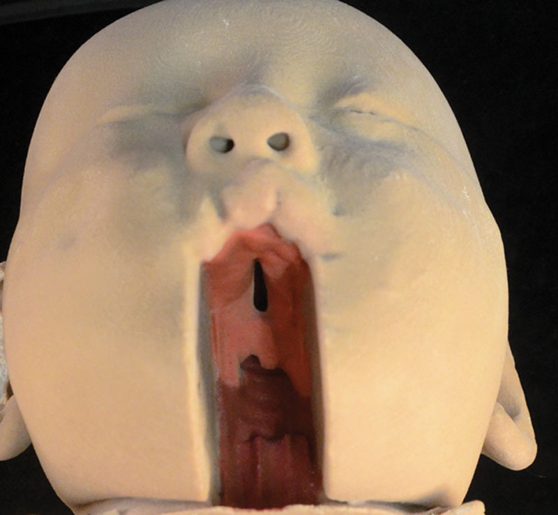
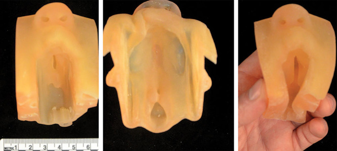
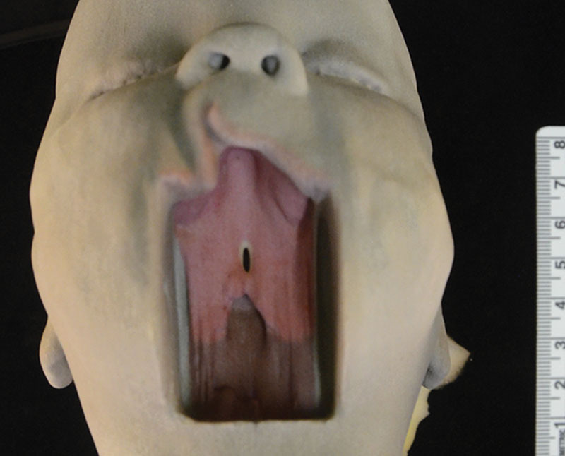
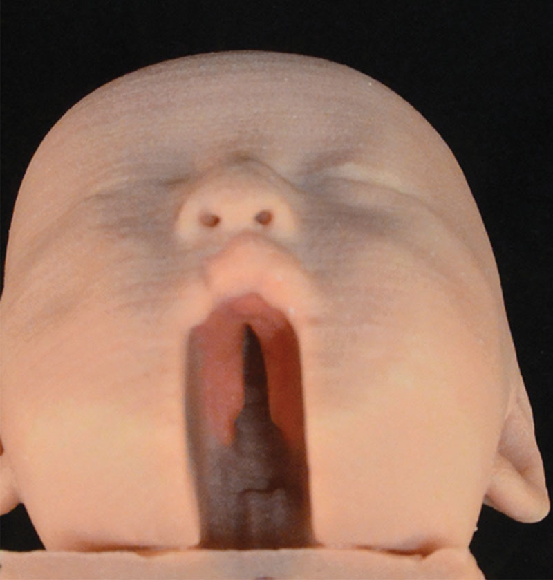
Magnetic resonance imaging data of 2 children (8 and 14 months) were segmented, colored, and anonymized, and stereolothographic files were prepared for 3D printing on either multicolor plastic or powder 3D printers and multimaterial 3D printers.
Two models were deemed of sufficient quality and anatomical accuracy to print unamended. One data set was further manipulated digitally to artificially extend the length of the cleft. Thus, 3 models were printed: 1 incomplete soft-palate deformity, 1 incomplete anterior palate deformity, and 1 complete cleft palate. All had cleft lip deformity. The single-material 3D prints are of sufficient quality to accurately identify the nature and extent of the deformities. Multimaterial prints were subsequently created, which could be valuable in surgical training.
Improvements in the quality and resolution of radiographic imaging combined with the advent of multicolor multiproperty printer technology will make it feasible in the near future to print 3D replicas in materials that mimic the mechanical properties and color of live human tissue making them potentially suitable for surgical training.
探讨腭裂畸形儿童3D打印模型的潜在可行性和局限性。
此前已描述过正常解剖标本3D打印复制品的优势。创建显示患者特异性解剖病理学的3D打印模型用于手术规划和干预是一个新兴领域。在此,我们探讨了利用罕见的儿科放射学数据集创建用于手术教育的3D打印模型的可能性。
对2名儿童(8个月和14个月)的磁共振成像数据进行分割、上色和匿名化处理,并准备立体光刻文件,以便在多色塑料或粉末3D打印机以及多材料3D打印机上进行3D打印。
有两个模型被认为质量和解剖准确性足以直接打印。对一个数据集进行了进一步的数字处理,人为延长了腭裂的长度。因此,打印了3个模型:1个不完全软腭裂畸形、1个不完全前腭裂畸形和1个完全腭裂。所有模型均伴有唇裂畸形。单材料3D打印的质量足以准确识别畸形的性质和程度。随后制作了多材料打印模型,这在手术训练中可能具有重要价值。
放射成像质量和分辨率的提高,再加上多色多属性打印机技术的出现,将使在不久的将来用模拟活体人体组织机械性能和颜色的材料打印3D复制品成为可能,使其有可能适用于手术训练。